Precise Imaging Expands with a New Radiology Center in Fresno, CA


The new center on West Shaw Lane features an Open MRI and X-Ray services, providing the community with enhanced diagnostic capabilities.


FRESNO, CA – Precise Imaging, a leading provider of diagnostic imaging services, is thrilled to announce the opening of its new radiology imaging center located at 2540 W. Shaw Lane, Suite 112-A, Fresno, CA 93711. The latest addition to the Precise Imaging network is now ready to serve the Fresno community and the surrounding areas, offering an Open MRI and X-Ray services.


With a continuous commitment to providing high-quality, accessible imaging services, Precise Imaging has invested in cutting-edge technology at its new Fresno center to ensure accurate and timely diagnoses. The Open MRI service aims to deliver a comfortable and less claustrophobic experience for patients, while the advanced X-Ray services ensure precise imaging for a variety of medical needs.


The Fresno location not only expands Precise Imaging’s geographical footprint but also reinforces its mission to offer patient-centric, technologically advanced imaging services to a broader spectrum of patients. This expansion underscores Precise Imaging's dedication to community healthcare and its endeavor to be at the forefront of diagnostic imaging services.


"We are elated to extend our services to the Fresno community," said Mike Rashidi, VP of Precise Imaging. "Our new center is equipped with an Open MRI and XRAY services to ensure precise and quick results. With a team of experienced radiologists and technologists, we are here to provide compassionate care and superior imaging services to all our patients."


The center is open M-F 8am-7pm and adheres to rigorous safety and sanitation protocols to ensure a safe environment for both patients and staff.


For more information about Precise Imaging and the new Fresno radiology imaging center, or to schedule an appointment, individuals can or call 800-558-2223


About Precise Imaging:


Precise Imaging offers a comprehensive range of diagnostic imaging services with a network of easily accessible imaging centers. With a focus on providing high-quality imaging and outstanding patient care, Precise Imaging is dedicated to meeting the diverse needs of patients, medical professionals, and healthcare organizations.


The Power of MRI Exams in Personal Injury Lawsuits


The health and wellbeing of individuals are often vulnerable to mishaps and accidents that can cause personal injuries. Such injuries could result in a legal dispute where the injured person, known as the plaintiff, seeks compensation for damages from the person or entity they believe caused their injury. In these instances, a comprehensive understanding of the injury's extent is paramount, often heavily relying on medical records and procedures. One such crucial diagnostic tool is the Magnetic Resonance Imaging (MRI) examination. This article delves into how MRI exams can provide substantial assistance in personal injury lawsuits.


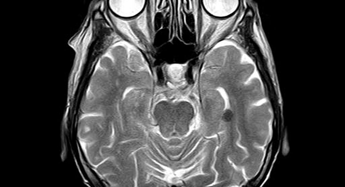
MRI is a non-invasive imaging technique that employs a powerful magnetic field and radio waves to generate detailed images of the inside of a body, typically focusing on soft tissues and organs. Unlike other imaging techniques like X-ray and CT scans, which primarily concentrate on skeletal injuries, MRI scans provide a comprehensive view of non-bony structures, like muscles, ligaments, tendons, and even the brain. This ability to visualize soft tissues makes MRI a valuable tool in evaluating the full extent of an injury.


In the context of personal injury lawsuits, MRI scans serve multiple critical roles. Primarily, it provides an objective basis for the plaintiff's claims of injury. Subjective symptoms like pain, discomfort, or restricted mobility may not convince a jury. However, visual evidence of damage, such as a torn ligament or herniated disc, becomes a tangible piece of evidence supporting the plaintiff's claims. By demonstrating the injury's existence and severity, MRI results can counter any argument that the plaintiff is exaggerating or feigning their condition.


Secondly, MRI results can contribute to determining the prognosis of an injury. The extent of damage revealed in the scan can help predict the duration of recovery, potential complications, and the possibility of permanent disability. This information is crucial in estimating the future medical costs, loss of earnings, and even non-economic damages like pain and suffering, which the plaintiff may include in their claim for compensation.


Furthermore, the timing of an MRI can also be a strategic element in a personal injury case. Early MRI exams might not reveal certain types of injuries like soft tissue damage, as these can take time to manifest. An MRI performed too soon could lead to an underestimation of the injury's extent, possibly harming the plaintiff's case. On the other hand, an MRI conducted too late might give the defendant an opportunity to argue that the injury was caused by something other than the initial incident. Hence, the timing of MRI is a critical factor in personal injury cases.


Finally, it's important to note that MRI results need to be interpreted and presented by a medical expert. Precise Imaging has several board certified radiologist that will review the images taken by the technologist and provide a report with their finding.


In conclusion, MRI examinations can be a powerful tool in personal injury lawsuits, aiding in confirming the existence and severity of an injury, predicting its prognosis, justifying medical expenses, and providing compelling evidence for the jury. However, these benefits are maximized when the timing of the MRI and interpretation of its results are strategically managed. It's crucial for individuals in these situations to consult with both medical and legal professionals to ensure the most effective use of MRI examinations in their case.


test tubes

What Are the Chances of an MRI Contrast Dye Allergy?

As we've covered in other articles, gadolinium-based contrast dyes play an essential role in magnetic resonance imaging (MRI) examinations. Contrast dyes allow technicians to capture high-quality images of various body structures, and for the most part, they're extremely well tolerated. Patients rarely suffer side effects from gadolinium, and when they do, those side effects are typically mild.

With that said, many patients have understandable concerns about magnetic resonance contrast media. If you've got a history of serious allergies, will gadolinium endanger your health?

To put it plainly, probably not. Hypersensitivity is extremely rare, and while patients should always discuss medical concerns with their physicians, we did some research to try to put gadolinium allergies in perspective.

If you’re not sure why gadolinium is necessary, be sure to read our article on contrast dyes.

How Common Are Gadolinium Allergies?


A 2012 study looked at 84,367 patients. Of those participants, 102 patients had hypersensitive reactions to gadolinium. That’s about 0.121 percent of the test group. If you’re otherwise healthy, the chances of an allergic reaction are extremely remote.

Other important takeaways from that study:

  • Women were more likely to have gadolinium allergies than men. Female patients had about 1.687 greater odds of suffering a hypersensitive reaction.

  • Patients with allergies were more likely to suffer a reaction. The odds ratio increased to about 2.829 for these patients, so if you have a history of asthma or allergies, be sure to tell your MRI team.

  • Patients who received multiple doses were more likely to suffer a reaction. If you regularly receive MRIs, you might eventually have a reaction, even if you haven’t had any side effects yet. Still, we’d like to hammer home this point: Reactions of any kind are extremely rare.

  • Most of those hypersensitive reactions weren’t severe. 91.1 percent of patients only suffered urticaria (the medical name for hives). A more severe anaphylactic reaction occurred in 11 cases (9.8 percent of allergic patients). Due to one fatality — and remember, this study looked at more than 84,000 patients — the mortality rate was 0.0007 percent.

  • Some dyes were more likely to cause a reaction than others. Gadodiamide, for instance, had a hypersensitivity rate of only 0.013 percent. That means that if you do have an allergy, your technicians can probably find a dye that you can tolerate.


When you set up your MRI, be sure to tell your physician and your imaging team about any allergies or any reactions you suffered in past procedures. However, don’t worry about the dye; it’s extremely safe, and even if you have a reaction, it will probably be treated easily before you leave the examination room.

Remember, if you need an MRI, you can save a tremendous amount of money by comparing costs. Precise Imaging has evaluated a large network of professional MRI clinics, and our convenient online tools help to make the process much less stressful. Call us at 800-558-2223 to book your appointment or email [email protected] to get started.
Metal Worker

The MRI for Metal Workers: Hazards and Solutions

Sheet metal workers, welders, and others exposed to tiny metal fragments face particular risks during an MRI scan. An adequate screening questionnaire will ask patients if they’ve been exposed to metal fragments well before they enter the MRI suite. In order to minimize anxiety both before and during imaging procedures, physicians should educate patients who work with metal early in the conversation.

Here are the key things to communicate to metal workers when referring them to a diagnostic imaging provider:

  • The presence of metal in the body may not present a health risk, but may still contraindicate MRI as an imaging modality.Even if metal fragments don’t react to the magnet in ways that can cause harm, they can still disrupt magnetic field homogeneity. This can cause visual artifacts and signal loss, limiting the diagnostic value of the resulting images.Senol and Gumus present a novel example of this distortion in a brief submission to the journal Quantitative Imaging in Medicine and Surgery. The patient they describe was a metal worker; despite the fact that he had showered and washed his hair prior to his MRI scan, some metal dust remained on his scalp. These fragments created strange circular objects, like water bubbles, on the resulting images. (These images were obtained on a 1.5 Tesla MRI scanner.)

  • Sheet metal workers are more likely than others to have tiny metal shards in their eyes, and these objects may not produce any symptoms at all.Patients are often unaware of the presence of intraocular foreign bodies. For instance, see this article from the American Journal of Ophthalmology Case Reports.Even if patients aren’t experiencing discomfort or pain, physicians may elect to obtain images of the eyes via nonmagnetic means before progressing to the MRI study.

  • Standard procedure is to order a CT orbit scan prior to MRI for patients who face higher risks of metal fragments in their eyes.CT scans don’t use magnets at all, and are safe for patients who have metal shavings in their eyes. The orbit CT scan is a quick, noninvasive way to make sure patients can safely receive an MRI in scanners of any strength.

  • Regardless of the findings of preliminary scanning, patients will always have a way to stop the MRI procedure for any reason.Patients will always have a route of communication with the attending technologist. If they have any concerns during the procedure, they can always tell their technologist, who will stop the scan and evaluate the situation before proceeding.

  • If the MRI scan poses any health threat at all, plenty of alternative imaging modalities are available to meet diagnostic goals.In the rare event that technologists and radiologists do find ferromagnetic metal fragments within the patient’s eyes or body, they can always use an alternative imaging technique. Scans involving X-rays don’t create magnetic fields, and won’t interact with metal implants or particles.


The pre-MRI screening process is designed to ensure safety for patients, and a big part of the effort is discerning the presence of ferromagnetic metallic objects within the body. The high-powered magnetic fields involved in an MRI scan can cause these objects to heat up, vibrate, or even shift location — clearly, this presents a health risk for patients, contraindicating MRI as a diagnostic imaging modality.

Despite these risks, physicians can help to provide a more comfortable treatment experience by discussing the above issues with qualifying patients from the beginning of the diagnostic process. Doctors can continue to order the MRI for metal workers with a high degree of confidence in the safety, efficacy, and comfort of their patients, and communication plays a central role in the process.

References:

Platt AS, Wajda BG, Ingram AD, Wei XC, Ells AL. Metallic intraocular foreign body as detected by magnetic resonance imaging without complications- A case report. Am J Ophthalmol Case Rep. 2017;7:76-79. Published 2017 Jun 22. doi:10.1016/j.ajoc.2017.06.010

Senol S, Gumus K. A rare incidence of metal artifact on MRI. Quant Imaging Med Surg. 2017;7(1):142-143.
Head MRI

MRI Scans with Contrast and How They Differ From MRI Scans Without Contrast

When recommending MRI examinations, physicians often encounter concerns from their patients—which is understandable, but largely unnecessary. Compared with other imaging technologies, MRIs provide high-quality images with very limited risks.

Even so, physicians should certainly understand the current science surrounding various gadolinium-based contrast dyes when making their recommendations. Gadolinium dyes provide enhanced clarity, which can be essential when making a diagnosis, but they’re not always necessary, and in some cases, they may be unsafe. Speak with a radiologist in order to make appropriate decisions on a case-by-case basis.

In this article, we’ll address several recent studies; note, however, that this is not intended as a comprehensive set of recommendations or as a thorough analysis of health risks.

What are the risks of gadolinium-based contrast agents?


Research shows that gadolinium can accumulate in the brain, most notably in the dentate nuclei and globus pallidus. As these regions of the brain are involved in motor function, it’s conceivable that motor function could be affected over time, particularly in patients who undergo numerous MRIs. Some forms of gadolinium may also disrupt the action of thyroid hormone, which is an important concern to keep in mind when administering MRIs to pregnant patients.

One perinatal in vivo study (performed on adult mice) found that:

… when gadoterate meglumine or gadodiamide was intravenously injected into dams during perinatal period (embryonic day 15–19, single injection/day), which is the critical period for the functional organization of neuronal circuits, both GBCAs disrupted motor coordination and impaired memory function [8]. The magnitude of disruption was higher with gadodiamide.

However, in human patients, these neurological effects haven’t been established. There’s no evidence to suggest that gadolinium deposition in the brain affects neurological function, and the mere presence of gadolinium deposits shouldn’t be perceived as a reason to forego dye-enhanced MRIs, particularly when dyes would provided a diagnostically useful image that wouldn’t be attainable otherwise.

A more pressing concern is nephrogenic systemic fibrosis, a rare condition which has been proposed as causally linked to gadolinium contrast dyes. This typically isn’t a concern in healthy patients, but gadolinium agents should not be administered to patients with severe kidney issues.

In the vast majority of cases, gadolinium contrast agents could allow for faster, more thorough treatment of serious health conditions, and the benefits of contrast dyes will greatly outweigh the potential risks.

Should physicians recommend contrast-free MRI scans to patients?


Since gadolinium deposits accumulate in the brain, contrast dyes shouldn’t be used haphazardly. A 2017 review from the International Society for Magnetic Resonance in Medicine (ISMRM) recommended that the MRI community avoid using gadolinium-based contrast agents when they are not necessary.

Of course, this is a fairly obvious conclusion for physicians—the real question is what constitutes a medical necessity. The ISMRM stopped short of recommending wide changes to the way that contrast dyes are used, and to date, there’s no evidence linking gadolinium deposits in the brain and adverse health effects.

It’s also important to note that different contrast dyes accumulate in different ways. While all gadolinium-based agents seem to form deposits in the aforementioned regions of the brain, the size of these deposits vary; gadoterate meglumine (macrocyclic GBCA), for instance, does not seem to significantly change thyroid hormone action.

With this in mind, physicians should recommending MRI scans with gadolinium-based contrast agents to pregnant women. When patients are likely to undergo numerous MRI scans, gadolinium-based contrast agents should be used as infrequently as possible.

Finally, patients who have a high susceptibility to renal failure should not be exposed to gadolinium-based contrast dyes, unless there is no viable alternative. When this is the case, lower-risk gadolinium agents are obviously preferable.

However, it’s important to note that, at this point, there is no evidence showing that gadolinium is inherently unsafe. When compared with the iodine-based agents used for x-ray examinations, gadolinium contrast dyes are largely preferable.

Physicians who need fast access to MRI services can make referrals through the Precise Imaging physician’s portal. This online tool provides anytime access to the crucial services we provide.

Call Precise Imaging at 800-558-2223 to make a referral or schedule an appointment today.

References:

Ariyani, W., Khairinisa, M.A., Perrotta, G. et al. The Effects of Gadolinium-Based Contrast Agents on the Cerebellum: from Basic Research to Neurological Practice and from Pregnancy to Adulthood. Cerebellum. (2018) 17: 247. https://doi.org/10.1007/s12311-017-0903-4

Goischke, HK. MRI With Gadolinium-Based Contrast Agents: Practical Help to Ensure Patient Safety. J Am Coll Radiol. 2016;13(8):890. https://doi.org/10.1016/j.jacr.2016.05.007

Gulani V, Calamante F, Shellock FG, Kanal E, Reeder SB. Gadolinium deposition in the brain: summary of evidence and recommendations. The Lancet. 2017;16(7):564-570. Published 2017 Jul 1. doi: doi.org/10.1016/S1474-4422(17)30158-8

Khairinisa MA et. al. The Effect of Perinatal Gadolinium-Based Contrast Agents on Adult Mice Behavior. Invest Radiol. 2018;53(2):110-118. Published Feb 2018. doi: 10.1097/RLI.0000000000000417

Schlaudecker JD, Bernheisel CR. Gadolinium-associated nephrogenic systemic fibrosis. Am Fam Physician. 2009:80(7):711-4. Published Oct 1 2009. PMID: 19817341.
Pacemaker

Medical Implants and MRI Scans: Updating the Research

Millions of potential MRI patients already live with a medical implant. The use of pacemakers has seen a more than 50 percent increase between 1993 and 2009, while the first nationwide study of knee and hip arthroplasties show that over 7.2 million Americans are living with implants.

These implants can increase mobility and improve a patient’s quality of life and life expectancy; there’s no question that they’re a net medical plus. But implants also come with new restrictions, including some that affect our use of MR technologies to obtain diagnostic images.

To be clear, MRI scans are incredibly helpful, low-risk, non-invasive imaging options. But sometimes those with medical implants aren’t able to take advantage of this imaging choice, and the list of affected implants is constantly in flux. That’s why it’s important for all physicians to stay up-to-date on the research regarding medical implants and MRI scans.

Understanding Interactions Between Medical Implants and MRI Scans


Because MRI scans use strong magnets, metal implants could potentially cause negative reactions or distort the images created during the test. Pacemakers, defibrillators, cochlear implants, or other small metal hardware can heat up or be drawn to the machine during an MRI. Electronic devices can also potentially malfunction during a scan, posing a risk to the patient.

That said, medical device manufacturers are always innovating. Increasingly, designers of these devices plan for MRI safety when they’re building their products. So where does the latest generation of medical implants stand when it comes to the safe use of MR modalities during the diagnostic process? That, of course, depends on the implant itself.

New Research on Common Medical Implants and MRI Scans


Pacemakers and defibrillators have long been on the MRI-unsafe list. Recent studies suggest that this abundance of caution is not necessary for most people. One piece in the Journal of Clinical Electrophysiology, labeled a “proof of concept study” by its authors, suggests that the benefits of conducting an MRI scan on patients with pacemakers and defibrillators justified the risk.

Meanwhile, a safety study of nearly 40 medical implants in a 7 Tesla MRI scanner found that only four of them — one heel implant, two stints, and one fibular implant — failed safety tests and earned the designation MR-unsafe for 7-T MRI usage.

Leading hospitals are also building the case for MRI procedures in patients with pacemakers and defibrillators. The Johns Hopkins Hospital has safely conducted MRI studies on more than 300 patients with cardiac implants at the time of this writing.

Of course, none of this is to say that all medical implants are always safe for MRI scans. But as the research continues, and as device manufacturers learn more about which materials and designs interact with MR fields and which don’t, it seems that the diagnostic power of MRI scans will become more and more available to patients who have implanted medical devices. That’s a good thing for patients and for medical science more broadly.
mri brain research

Diagnosing Traumatic Brain Injury: CT Scans and MRI Tests


Researchers suspect that the true rate of mild traumatic brain injuries remains much higher than reported, suggesting the need for broader awareness of the condition. Diagnosis through imaging studies can help.


Neuroradiologists possess powerful tools for revealing the presence of traumatic brain injury (TBI): the CT scan and the MRI scan. Not every patient with suspected TBI requires imaging studies, but for those who do, these two techniques can greatly improve outcomes through prompt diagnosis.


Each of these diagnostic techniques carries its own strengths and hazards, and physicians order them in different cases. A thorough understanding of brain imaging studies can help the medical community identify cases of TBI more readily, leading to better, faster interventions.    


This is a subject of increasing concern among doctors; traumatic brain injuries — especially of the mild variety, more commonly known as concussions — are fairly common. In 2013, around 2.5 million people visited an emergency room with TBI-related complaints. Most cases in the 15-24 age range are related to motor vehicle accidents, but among causes, there’s a close second: playing sports.   


Measuring TBI in the Sports Community


The sports community is particularly interested in improving treatment for TBI, and for good reason. Remember that more concussions and brain injuries come from playing sports than any other cause except for car accidents.


A brief glimpse through the numbers paints an alarming picture:


These statistics explain why researchers are working so hard to find fast, field-side imaging tests that can pinpoint the severity of TBI immediately following the event. Promising options include stadium MRI rooms and highly portable ultrasound; still, for most players at all levels, radiology-assisted diagnosis of TBI will involve a trip to the imaging center. This places us firmly back in CT/MRI territory.  


CT Scans in the Diagnosis of TBI


Computerized tomography (CT) scans take multiple X-rays and combine them into cross-sectional “slices” of internal structures. While this exposes the patient to small doses of ionizing radiation, but it’s also the fastest, most accurate way to identify bleeding and swelling in the brain.


Clearly, edema (brain swelling) and hematoma (bleeding in and/or around the brain) are serious conditions. The faster doctors discover them, the better for the patient. Typically, then, physicians order CT scans for suspected acute injuries to the brain. The CT scan is the modality of first access.


Later, doctors may order more CT scans to track healing in TBI instances that don’t require surgery. As with every order of an X-ray procedure, physicians weigh the benefits of the treatment against the risks posed by exposure to radiation before making a referral.  


MRI Scans in the Diagnosis of TBI


Magnetic resonance imaging (MRI) is the go-to tool for identifying subtle effects of injury, including bruising, scarring, and microscopic damage to nerve fibers. Images produced by CT scan won’t reveal these conditions, though nerve fiber injury is a common cause of stubborn symptoms.


Sometimes brain tissue is injured too severely to recover, so MRI scans can track the results of a TBI for years following the precipitating event. This imaging modality is another powerful tool in the neuroradiologist’s brain-injury kit.  


Schedule Diagnostic Imaging for Patients with TBI


Ultimately, health care providers will determine the appropriate imaging technique for each patient showing signs of TBI. Plenty of non-radiological tests exist; these may be enough to recognize and begin treatment for milder injuries to the brain.


Physicians who need fast access to radiology services in cases of TBI can make referrals through the Precise Imaging physician’s portal. This online tool provides anytime access to the crucial services we provide.


Call Precise Imaging at 800-558-2223 to make a referral or schedule an appointment today.


References:


Centers for Disease Control and Prevention Justification of Estimates for Appropriation Committees, Fiscal Year 2016.” CDC. Centers for Disease Control and Prevention, U.S Department of Health and Human Services, 2016. PDF. 2 Jan. 2019.


Orenstein, Beth. “A Closer Look at Concussions.RadiologyToday. Great Valley Publishing Company, Inc., Sept. 2016. Web. 2 Jan. 2019.


Prince, Carolyn and Maya Bruhns. “Evaluation and Treatment of Mild Traumatic Brain Injury: The Role of Neuropsychology.NLM. Brain Sciences, Aug. 2017. Web. 2 Jan. 2019.


Traumatic Brain Injury (TBI) and Concussion.ASNR. American Society of Neuroradiology, n.d. Web. 2 Jan. 2019.


Comparing MRI and CT Scans in Personal Injury Cases

Comparing MRI and CT Scans in Personal Injury Cases


Diagnostic imaging is crucial to many personal injury lawsuits, but what type of imaging do your clients need? Odds are, doctors and legal teams will point toward one of two options: an MRI scan or a CT scan.


These procedures could provide crucial evidence in your case, clarifying to jurors and the judge what was merely conjecture before. Here are the main differences between CT scans and MRI scans, along with a few things personal injury attorneys should know about these popular diagnostic imaging modalities.

CT Scans and How They Work


Computerized Axial Tomography, otherwise known as a CT scan, uses X-rays to produce detailed images of the human body. The basic concept is this: The machine shoots a narrow beam through the target area of the body. Then it rotates the beam, creating a cross-sectional image.


Most of the radiation passes through the body, but when it hits denser material such as bone, it stops. This is how bones show up on the screen as a contrast. Through the use of multiple images stacked on top of each other, the computer program creates a 3D picture of the patients insides.  


The scan is a non-invasive procedure. Note, however, that it does expose the patient to ionizing radiation.      

MRI Scans and How They Work


Magnetic Resonance Imaging, or MRI, uses powerful magnets to create a strong magnetic field. The machine then sends pulses through the patient’s tissues to create clear 3D images of the target area.


This technology does use damaging radiation during photo capture, and it’s totally non-invasive. The patient rests inside a large magnet during the procedure, and must remain quite still during the whole process, or else risk a distorted result. However, resulting images are often highly accurate and detailed.


The Benefits of MRI Scans and CT Scans in Personal Injury Cases


If a picture is really worth a thousand words, in the courtroom, it could be worth many thousands of dollars. Some sort of diagnostic imaging can strengthen virtually any case.


Of course, each imaging modality offers specific benefits to the patient and client, depending on the nature of the injury itself. CT scans, for example, are far better at diagnosing breaks, fractures, and other types of bone damage.


Due to a lack of water in bones, a necessary component to MRI imaging, they don’t provide a lot of detail in MRI images. On the other hand, MRI scans are excellent at imaging soft tissues, like ligaments, muscles, tendons and nerves.

The Disadvantages of CT Scans and MRI Scans in Establishing Injury


Both of these image modalities have disadvantages, although not serious ones. CT scans sometimes do use contrasting agents, so make sure there is no allergic reaction history before agreeing to this aspect of the procedure. CT scans also use X-rays to produce image, although only those who are pregnant should avoid them as a result and even then, only the areas of the abdomen and pelvis should be avoided.


In regards to an MRI, due to the strong magnetic field used, people with certain implants (especially those with iron, such as a pacemaker), should never enter an MRI machine. The MRI is also quite loud, and in certain cases could induce claustrophobia, so if there is a patient history of this clinical diagnoses should generally avoid MRI scans.


The main takeaway here is that both CT and MRI scans can provide tremendous legal benefits during a personal injury case. They may provide powerful evidence of the extent of injury. Of course, attorneys should take their lead from doctors. The leading factor in deciding which image modality is determining the type of trauma after consultation with a medical professional.

Gadolinium Retention Research Roadmap Now Available

Gadolinium Retention Research Roadmap Now Available


On February 15, 2018, The National Institute of Biomedical Imaging and Bioengineering brought together an international group of researchers, scientists, and medical doctors to look at the current and future concerns regarding the human body’s retention of gadolinium-based contrast agents (GBCAs). Ultimately, they wished to identify any potential safety hazards associated with this common MRI contrast agent. In September, the results were published, mapping out a path for future research into this subject. Physicians can now access the work for a greater understanding of GBCAs used in diagnostic imaging settings.   


The report states that “in spite of more than 30 years of use of GBCAs, important information about the biodistribution and tissue interactions of each GBCA in clinical use remains unknown. It is clear that gadolinium retention in a number of tissues, including bone, skin, and brain, beyond 24 hours may occur with all types of GBCAs, although the magnitude of observed retention is greater with linear GBCAs than with macrocyclic GBCAs.”

What Researchers Know About GBCAs Today


More simply put, the researchers found the body does have a tendency to retain GCBAs for more than 24 hours after injection, although the degree to which they are retained depends upon the GBCA type as well as the specific organ. Another finding by the authors of the report essentially states that much more research is needed to determine more exactly how much the body retains GCBAs, and what, if any, safety risks accompany this retention.


Despite an estimated 450 million or more intravenous doses of GCBAs, very little is understood regarding their potential health consequences. While the substance is largely regarded as safe, some types of GCBAs have been linked to nephrogenic systemic fibrosis in patients with advanced forms of kidney diseases. This is why the use of GCBAs is usually contraindicated for patients with serious kidney problems.


The GBCA Research Roadmap Plots a Course Forward for Researchers


The new roadmap illustrates key concerns and identifies certain subsets of the population that need to be more closely studied. Populations of study include pregnant women, the elderly, younger patients, and lactating women. Research efforts should be aided by utilizing large database sets such as those at the Mayo Clinic, or studies in which patients underwent contrast aided imaging using GCBAs. Use of large amounts of data could help to uncover inconsistencies or particularly vulnerable groups when it comes to GCBA retention.   


The study acknowledges that future research will bridge important gaps in knowledge. The roadmap generated by the researchers prioritizes discovering  “(a) if gadolinium retention adversely affects the function of human tissues, (b) if retention is causally associated with short- or long-term clinical manifestations of disease, and (c) if vulnerable populations, such as children, are at greater risk for experiencing clinical disease.”


While this new document does not offer any answers to these questions, it does lay the groundwork for future research efforts in order to fill in these gaps. In fact, by plainly stating what is not known regarding GCBA retention, the authors of the “roadmap” have already taken an important first step.  


Access the full text of this special report here.


How Digital Access to Medical Images Helps Personal Injury Lawyers

How Digital Access to Medical Images Helps Personal Injury Lawyers


Diagnostic imaging is one of the clearest, most powerful ways to demonstrate objective manifestation of injury in a lawsuit. But like any piece of evidence in trial law, quality matters. Standard-quality images that clearly show a broken bone or inflammation to a trained radiologist might be meaningless to the jury. Medical images in the legal realm must be pristine.  


Luckily, we've come a long way from the days of lightboxes in the courtroom. Precise Imaging offers attorneys 24/7 access to medical images in their cases through a dedicated portal built just for them. These digital files provide a number of advantages that can make it much easier than it used to be to prove objective manifestation. Here are just a few of these benefits:


  • Digital images are easy to display in high-definition in a variety of settings. High-definition digital images look great whether you're working with an iPad on the train or displaying on a large-screen TV in front of the entire courtroom. You can also send them in an instant, rather than relying on physical CDs or film.


  • Cloud access allows attorneys to work when and where they must. Cloud storage keeps reports and images themselves available for working remotely. Lawyers simply log into the Precise Imaging attorneys portal to view their clients' medical imaging results at any time of the day or night. Even the tech support is available 24/7, ensuring the sort of flexibility today's personal injury professionals need.  


  • Collaboration is easier when all parties can remotely view the same images and reports. It isn't always easy to get the whole team together in a room. The cloud makes it possible for attorneys, their clients, and their legal teams collaborate remotely by viewing images together, at the same time, but in different places.


  • Digital files are easy to share with opposing counsel during discovery. The plaintiff's team isn't the only ones who need access to medical images. When it comes time to share this critical data with the opposing counsel, digital access makes things easy. The remote characteristics of cloud storage let everyone appropriate — including opposing counsel — access medical images when they must. Attorney's web portals through Precise Imaging streamline the process.    


The Precise Imaging attorney's portal is just one of the ways we serve attorneys. We also accept letters of protection and liens, whether they're associated with personal injury suits or workers' compensation cases.


Perhaps most importantly, we have the capacity to handle an attorney's entire case load with a single point of contact. Rather than searching through multiple log-ins and customer-service numbers for imaging services, attorneys who work with Precise Imaging can keep all of their case data at hand, with HIPAA-compliant security.


To learn more, or to refer a client, call Precise Imaging at 800-558-2223.