Comparing MRI and CT Scans in Personal Injury Cases
Comparing MRI and CT Scans in Personal Injury Cases
Diagnostic imaging is crucial to many personal injury lawsuits, but what type of imaging do your clients need? Odds are, doctors and legal teams will point toward one of two options: an MRI scan or a CT scan.
These procedures could provide crucial evidence in your case, clarifying to jurors and the judge what was merely conjecture before. Here are the main differences between CT scans and MRI scans, along with a few things personal injury attorneys should know about these popular diagnostic imaging modalities.
CT Scans and How They Work
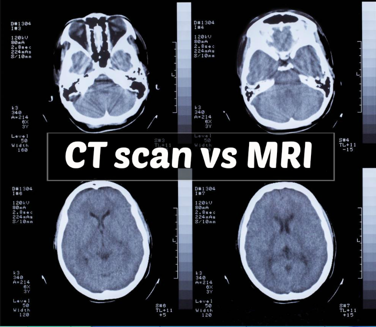
Computerized Axial Tomography, otherwise known as a CT scan, uses X-rays to produce detailed images of the human body. The basic concept is this: The machine shoots a narrow beam through the target area of the body. Then it rotates the beam, creating a cross-sectional image.
Most of the radiation passes through the body, but when it hits denser material such as bone, it stops. This is how bones show up on the screen as a contrast. Through the use of multiple images stacked on top of each other, the computer program creates a 3D picture of the patients insides.
The scan is a non-invasive procedure. Note, however, that it does expose the patient to ionizing radiation.
MRI Scans and How They Work
Magnetic Resonance Imaging, or MRI, uses powerful magnets to create a strong magnetic field. The machine then sends pulses through the patient’s tissues to create clear 3D images of the target area.
This technology does use damaging radiation during photo capture, and it’s totally non-invasive. The patient rests inside a large magnet during the procedure, and must remain quite still during the whole process, or else risk a distorted result. However, resulting images are often highly accurate and detailed.
The Benefits of MRI Scans and CT Scans in Personal Injury Cases
If a picture is really worth a thousand words, in the courtroom, it could be worth many thousands of dollars. Some sort of diagnostic imaging can strengthen virtually any case.
Of course, each imaging modality offers specific benefits to the patient and client, depending on the nature of the injury itself. CT scans, for example, are far better at diagnosing breaks, fractures, and other types of bone damage.
Due to a lack of water in bones, a necessary component to MRI imaging, they don’t provide a lot of detail in MRI images. On the other hand, MRI scans are excellent at imaging soft tissues, like ligaments, muscles, tendons and nerves.
The Disadvantages of CT Scans and MRI Scans in Establishing Injury
Both of these image modalities have disadvantages, although not serious ones. CT scans sometimes do use contrasting agents, so make sure there is no allergic reaction history before agreeing to this aspect of the procedure. CT scans also use X-rays to produce image, although only those who are pregnant should avoid them as a result and even then, only the areas of the abdomen and pelvis should be avoided.
In regards to an MRI, due to the strong magnetic field used, people with certain implants (especially those with iron, such as a pacemaker), should never enter an MRI machine. The MRI is also quite loud, and in certain cases could induce claustrophobia, so if there is a patient history of this clinical diagnoses should generally avoid MRI scans.
The main takeaway here is that both CT and MRI scans can provide tremendous legal benefits during a personal injury case. They may provide powerful evidence of the extent of injury. Of course, attorneys should take their lead from doctors. The leading factor in deciding which image modality is determining the type of trauma after consultation with a medical professional.