Precise Imaging Launches Innovative ‘Call Timing’ and ‘Call Me’ Feature for Enhanced Customer Convenience
Agoura Hills, CA – 01/30/2024 – Precise Imaging, a leader in diagnostic imaging services, is proud to announce the launch of a revolutionary new feature on its website: "When to Call" & "Call Me". This innovative service is designed to significantly enhance the convenience for patients, referring physicians, and attorneys who use Precise Imaging's services.
The new "When to Call" feature on the Precise Imaging website addresses a common challenge in healthcare communication: finding the best time to call for information or assistance. Recognizing this issue, Precise Imaging has developed a dual-component solution to streamline communication and improve user experience.
You can use the released feature by visiting this link: https://www.precisemri.com/when-to-call
Key Features of "When to Call":
- Best Time to Call Indicator: This aspect of the service informs users of the most optimal times to call Precise Imaging, reducing wait times and ensuring more efficient communication. By guiding users to contact at less busy hours, the feature promises a more seamless and satisfying interaction with Precise Imaging’s dedicated customer service team.
- Call Me Option: Understanding that time is precious, Precise Imaging offers a 'Call Me' option. With this, users can simply request a call back from the service team. This means they no longer need to spend time waiting on the line. Instead, Precise Imaging will call them back as soon as they are first in line, ensuring no unnecessary waiting and a more personalized service experience.
A Commitment to Customer Satisfaction:
"At Precise Imaging, we are constantly seeking ways to enhance our service and ensure our clients have the best possible experience," said Danny Rackow, IT Director, of Precise Imaging. "The 'Call Me' feature is a testament to our commitment to customer convenience and satisfaction. We understand the value of our clients' time and are dedicated to making our services as accessible and user-friendly as possible."
This latest feature is part of Precise Imaging's ongoing effort to leverage technology for improved customer service in the healthcare sector. With a focus on innovation and client satisfaction, Precise Imaging continues to set the standard for excellence in diagnostic imaging services.
About Precise Imaging:
Precise Imaging is a leading provider of diagnostic imaging services, with a network of facilities across the United States. Known for its state-of-the-art equipment, highly trained staff, and commitment to quality patient care, Precise Imaging is at the forefront of the medical imaging industry.
Precise Imaging Expands with a New Radiology Center in Fresno, CA
The new center on West Shaw Lane features an Open MRI and X-Ray services, providing the community with enhanced diagnostic capabilities.
FRESNO, CA – Precise Imaging, a leading provider of diagnostic imaging services, is thrilled to announce the opening of its new radiology imaging center located at 2540 W. Shaw Lane, Suite 112-A, Fresno, CA 93711. The latest addition to the Precise Imaging network is now ready to serve the Fresno community and the surrounding areas, offering an Open MRI and X-Ray services.
With a continuous commitment to providing high-quality, accessible imaging services, Precise Imaging has invested in cutting-edge technology at its new Fresno center to ensure accurate and timely diagnoses. The Open MRI service aims to deliver a comfortable and less claustrophobic experience for patients, while the advanced X-Ray services ensure precise imaging for a variety of medical needs.
The Fresno location not only expands Precise Imaging’s geographical footprint but also reinforces its mission to offer patient-centric, technologically advanced imaging services to a broader spectrum of patients. This expansion underscores Precise Imaging's dedication to community healthcare and its endeavor to be at the forefront of diagnostic imaging services.
"We are elated to extend our services to the Fresno community," said Mike Rashidi, VP of Precise Imaging. "Our new center is equipped with an Open MRI and XRAY services to ensure precise and quick results. With a team of experienced radiologists and technologists, we are here to provide compassionate care and superior imaging services to all our patients."
The center is open M-F 8am-7pm and adheres to rigorous safety and sanitation protocols to ensure a safe environment for both patients and staff.
For more information about Precise Imaging and the new Fresno radiology imaging center, or to schedule an appointment, individuals can or call 800-558-2223
About Precise Imaging:
Precise Imaging offers a comprehensive range of diagnostic imaging services with a network of easily accessible imaging centers. With a focus on providing high-quality imaging and outstanding patient care, Precise Imaging is dedicated to meeting the diverse needs of patients, medical professionals, and healthcare organizations.

The MRI for Metal Workers: Hazards and Solutions
Here are the key things to communicate to metal workers when referring them to a diagnostic imaging provider:
- The presence of metal in the body may not present a health risk, but may still contraindicate MRI as an imaging modality.Even if metal fragments don’t react to the magnet in ways that can cause harm, they can still disrupt magnetic field homogeneity. This can cause visual artifacts and signal loss, limiting the diagnostic value of the resulting images.Senol and Gumus present a novel example of this distortion in a brief submission to the journal Quantitative Imaging in Medicine and Surgery. The patient they describe was a metal worker; despite the fact that he had showered and washed his hair prior to his MRI scan, some metal dust remained on his scalp. These fragments created strange circular objects, like water bubbles, on the resulting images. (These images were obtained on a 1.5 Tesla MRI scanner.)
- Sheet metal workers are more likely than others to have tiny metal shards in their eyes, and these objects may not produce any symptoms at all.Patients are often unaware of the presence of intraocular foreign bodies. For instance, see this article from the American Journal of Ophthalmology Case Reports.Even if patients aren’t experiencing discomfort or pain, physicians may elect to obtain images of the eyes via nonmagnetic means before progressing to the MRI study.
- Standard procedure is to order a CT orbit scan prior to MRI for patients who face higher risks of metal fragments in their eyes.CT scans don’t use magnets at all, and are safe for patients who have metal shavings in their eyes. The orbit CT scan is a quick, noninvasive way to make sure patients can safely receive an MRI in scanners of any strength.
- Regardless of the findings of preliminary scanning, patients will always have a way to stop the MRI procedure for any reason.Patients will always have a route of communication with the attending technologist. If they have any concerns during the procedure, they can always tell their technologist, who will stop the scan and evaluate the situation before proceeding.
- If the MRI scan poses any health threat at all, plenty of alternative imaging modalities are available to meet diagnostic goals.In the rare event that technologists and radiologists do find ferromagnetic metal fragments within the patient’s eyes or body, they can always use an alternative imaging technique. Scans involving X-rays don’t create magnetic fields, and won’t interact with metal implants or particles.
The pre-MRI screening process is designed to ensure safety for patients, and a big part of the effort is discerning the presence of ferromagnetic metallic objects within the body. The high-powered magnetic fields involved in an MRI scan can cause these objects to heat up, vibrate, or even shift location — clearly, this presents a health risk for patients, contraindicating MRI as a diagnostic imaging modality.
Despite these risks, physicians can help to provide a more comfortable treatment experience by discussing the above issues with qualifying patients from the beginning of the diagnostic process. Doctors can continue to order the MRI for metal workers with a high degree of confidence in the safety, efficacy, and comfort of their patients, and communication plays a central role in the process.
References:
Platt AS, Wajda BG, Ingram AD, Wei XC, Ells AL. Metallic intraocular foreign body as detected by magnetic resonance imaging without complications- A case report. Am J Ophthalmol Case Rep. 2017;7:76-79. Published 2017 Jun 22. doi:10.1016/j.ajoc.2017.06.010
Senol S, Gumus K. A rare incidence of metal artifact on MRI. Quant Imaging Med Surg. 2017;7(1):142-143.

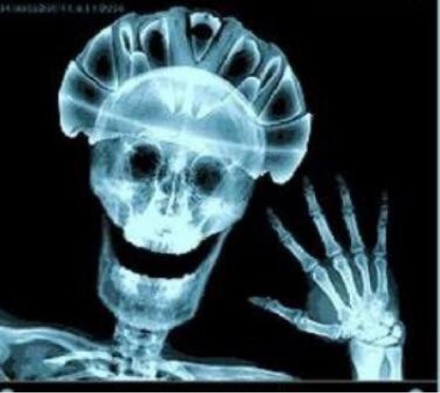
Diagnosing Traumatic Brain Injury: CT Scans and MRI Tests
Researchers suspect that the true rate of mild traumatic brain injuries remains much higher than reported, suggesting the need for broader awareness of the condition. Diagnosis through imaging studies can help.
Neuroradiologists possess powerful tools for revealing the presence of traumatic brain injury (TBI): the CT scan and the MRI scan. Not every patient with suspected TBI requires imaging studies, but for those who do, these two techniques can greatly improve outcomes through prompt diagnosis.
Each of these diagnostic techniques carries its own strengths and hazards, and physicians order them in different cases. A thorough understanding of brain imaging studies can help the medical community identify cases of TBI more readily, leading to better, faster interventions.
This is a subject of increasing concern among doctors; traumatic brain injuries — especially of the mild variety, more commonly known as concussions — are fairly common. In 2013, around 2.5 million people visited an emergency room with TBI-related complaints. Most cases in the 15-24 age range are related to motor vehicle accidents, but among causes, there’s a close second: playing sports.
Measuring TBI in the Sports Community
The sports community is particularly interested in improving treatment for TBI, and for good reason. Remember that more concussions and brain injuries come from playing sports than any other cause except for car accidents.
A brief glimpse through the numbers paints an alarming picture:
- In 2009, the Centers for Disease Control and Prevention estimate that nearly 250,000 children and teenagres visit emergency departments for concussions received while playing sports.
- The reported risk of concussion among football players is as high as 75 percent, reports Radiology Today, citing the Sports Concussion Institute.
- The University of Pittsburgh’s Brain Trauma Research Center estimates that about 300,000 concussions are associated with sports in a given year.
- The same institution lists the chance of concussion for athletes in contact sports to be up to 19 percent per year of play.
These statistics explain why researchers are working so hard to find fast, field-side imaging tests that can pinpoint the severity of TBI immediately following the event. Promising options include stadium MRI rooms and highly portable ultrasound; still, for most players at all levels, radiology-assisted diagnosis of TBI will involve a trip to the imaging center. This places us firmly back in CT/MRI territory.
CT Scans in the Diagnosis of TBI
Computerized tomography (CT) scans take multiple X-rays and combine them into cross-sectional “slices” of internal structures. While this exposes the patient to small doses of ionizing radiation, but it’s also the fastest, most accurate way to identify bleeding and swelling in the brain.
Clearly, edema (brain swelling) and hematoma (bleeding in and/or around the brain) are serious conditions. The faster doctors discover them, the better for the patient. Typically, then, physicians order CT scans for suspected acute injuries to the brain. The CT scan is the modality of first access.
Later, doctors may order more CT scans to track healing in TBI instances that don’t require surgery. As with every order of an X-ray procedure, physicians weigh the benefits of the treatment against the risks posed by exposure to radiation before making a referral.
MRI Scans in the Diagnosis of TBI
Magnetic resonance imaging (MRI) is the go-to tool for identifying subtle effects of injury, including bruising, scarring, and microscopic damage to nerve fibers. Images produced by CT scan won’t reveal these conditions, though nerve fiber injury is a common cause of stubborn symptoms.
Sometimes brain tissue is injured too severely to recover, so MRI scans can track the results of a TBI for years following the precipitating event. This imaging modality is another powerful tool in the neuroradiologist’s brain-injury kit.
Schedule Diagnostic Imaging for Patients with TBI
Ultimately, health care providers will determine the appropriate imaging technique for each patient showing signs of TBI. Plenty of non-radiological tests exist; these may be enough to recognize and begin treatment for milder injuries to the brain.
Physicians who need fast access to radiology services in cases of TBI can make referrals through the Precise Imaging physician’s portal. This online tool provides anytime access to the crucial services we provide.
Call Precise Imaging at 800-558-2223 to make a referral or schedule an appointment today.
References:
“Centers for Disease Control and Prevention Justification of Estimates for Appropriation Committees, Fiscal Year 2016.” CDC. Centers for Disease Control and Prevention, U.S Department of Health and Human Services, 2016. PDF. 2 Jan. 2019.
Orenstein, Beth. “A Closer Look at Concussions.” RadiologyToday. Great Valley Publishing Company, Inc., Sept. 2016. Web. 2 Jan. 2019.
Prince, Carolyn and Maya Bruhns. “Evaluation and Treatment of Mild Traumatic Brain Injury: The Role of Neuropsychology.” NLM. Brain Sciences, Aug. 2017. Web. 2 Jan. 2019.
“Traumatic Brain Injury (TBI) and Concussion.” ASNR. American Society of Neuroradiology, n.d. Web. 2 Jan. 2019.

Digital Sharing for Medical Images: PACS vs. VNAs
Health care providers are virtually required to have digital infrastructure that includes sharable medical images these days. But if you’re looking to upgrade your system, or you’re implementing your first digital image archive, you have many software products to choose from.
The leading technologies for storing and retrieving medical imaging files are picture archiving and communications systems (PACS) and vendor neutral archives (VNAs). While the two competing formats look similar on first glance, there are notable differences between them. Before we get to the contrasts, though, it’s important to note what PACS and VNAs have in common. Here are a few of the constants across both types of imaging technologies:
- Both PACS and VNAs provide remote access to images. Radiologists can upload images on one terminal, while physicians in a different office can log in to access them. This is the crucial requirement of all digital image-sharing systems.
- Both systems operate with the same file format and transmission protocol: DICOM, which stands for “digital imaging and communications in medicine.” File formats can lead to accessibility problems, as each application is only equipped to handle certain types of files. The universal use of DICOMs between PACS and VNAs seems to suggest that migrating images between the two systems would be seamless. Unfortunately, that’s rarely the case. We’ll get into that a little later.
- Both options may or may not also operate as a platform for non-DICOM images. This is an important question to ask when considering changes to an image-sharing system for medical care. If departments other than radiology are hoping to upload and access images through the same system, it is vital to choose software that can support non-DICOM images.
- Designers of both PACS and VNAs are working to improve mobility and access to images across different types of devices. That’s one of the leading requests by radiologists and other health care providers — physicians want to be able to call up an image, safely and with full HIPAA compliance, on a tablet or even a phone in the examination room with their patients. They also want access at office desktops for consultation and reporting.
Despite these similarities, PACS and VNAs have very different sets of advantages. To complicate matters, many PACS are beginning to provide some of the features that initially launched the popularity of the VNA. Before making a system-wide purchase (or subscription service, which is also available from many vendors), it’s important to consult all stakeholders in your health care system, and to get to know your vendor and their product well.
That said, there are some broad-strokes difference between your average PACS and a typical VNA. We’ll get into those next.
PACS vs. VNAs: Differences Between Medical Image-Sharing Platforms
According to the trade publication Diagnostic Imaging, the VNA tends to focus on archiving and backing up data, while the PACS usually emphasizes workflow and user experience. Of course, these general principles are less significant in today’s market, where there are a wide variety of VNAs and PACS, and their strong points have begun to overlap.
That said, here are some of the differences between PACS and VNAs that users and medical industry analysts have pointed out:
- The PACS is the original technology used to archive and retrieve digital medical images. As such, it’s often the choice of individual radiology departments, which were among the first to adopt digital imaging. VNAs, on the other hand, are more often found in multiple departments.
- PAC systems are more highly proprietary than most VNAs. That is, each individual PACS will require its own user interface and its own log-in information. VNAs were designed to support storage and access across systems and vendors. Of course, new developments in PACS technology renders this difference conditional, but it remains the conventional wisdom among medical technology experts.
- A VNA, by definition, divorces the storage/access functions of a PAC from a particular workstation or data silo. It uses its own application engine, allowing users to access images from multiple sources with the same user interface. That’s what makes VNAs typically better for interoperability between systems compared to a traditional PACS.
While the above list does seem to argue for the dominance of VNAs over PACS, in actual practice, implementation isn’t always the best choice for every provider. It can be expensive and time-consuming to migrate data from an existing PACS to a brand new VNA.
That’s because DICOM files contain both metadata and location pointers. The former attached patient information to the image; the latter helps the system find and pull up the specific image the user searches for.
During transition, all of this supplemental information can become scrambled, preventing access to images. In order to avoid this outcome, vendors often must reset DICOM headers and location pointers to ensure accuracy and access in the new system. That can be a lengthy and cost-intensive process.
Ultimately, then, institutions with the time and the money will benefit from an upgraded VNA system. More practically, some users will choose a PACS, with or without newly developed features. Regardless of the archiving and retrieval system, digital transmission for diagnostic images is practically a requirement in today’s medical system. Choose an imaging software product that makes it easy to share images with other departments and institutions.
References:
Jackson, Whitney. “What You Need to Know About VACS and VNA.” DiagnosticImaging.” UBM, 4 Sept. 2014. Web. 15 Oct. 2018.
O’Dowd, Elizabeth. “Pros and Cons of PACS, VNAs for Medical Image Data Storage.” HitInfrastructure. Xtelligent Healthcare Media, LLC, n.d. Web. 15 Oct. 2018.
Gadolinium Retention Research Roadmap Now Available
On February 15, 2018, The National Institute of Biomedical Imaging and Bioengineering brought together an international group of researchers, scientists, and medical doctors to look at the current and future concerns regarding the human body’s retention of gadolinium-based contrast agents (GBCAs). Ultimately, they wished to identify any potential safety hazards associated with this common MRI contrast agent. In September, the results were published, mapping out a path for future research into this subject. Physicians can now access the work for a greater understanding of GBCAs used in diagnostic imaging settings.
The report states that “in spite of more than 30 years of use of GBCAs, important information about the biodistribution and tissue interactions of each GBCA in clinical use remains unknown. It is clear that gadolinium retention in a number of tissues, including bone, skin, and brain, beyond 24 hours may occur with all types of GBCAs, although the magnitude of observed retention is greater with linear GBCAs than with macrocyclic GBCAs.”
What Researchers Know About GBCAs Today
More simply put, the researchers found the body does have a tendency to retain GCBAs for more than 24 hours after injection, although the degree to which they are retained depends upon the GBCA type as well as the specific organ. Another finding by the authors of the report essentially states that much more research is needed to determine more exactly how much the body retains GCBAs, and what, if any, safety risks accompany this retention.
Despite an estimated 450 million or more intravenous doses of GCBAs, very little is understood regarding their potential health consequences. While the substance is largely regarded as safe, some types of GCBAs have been linked to nephrogenic systemic fibrosis in patients with advanced forms of kidney diseases. This is why the use of GCBAs is usually contraindicated for patients with serious kidney problems.
The GBCA Research Roadmap Plots a Course Forward for Researchers
The new roadmap illustrates key concerns and identifies certain subsets of the population that need to be more closely studied. Populations of study include pregnant women, the elderly, younger patients, and lactating women. Research efforts should be aided by utilizing large database sets such as those at the Mayo Clinic, or studies in which patients underwent contrast aided imaging using GCBAs. Use of large amounts of data could help to uncover inconsistencies or particularly vulnerable groups when it comes to GCBA retention.
The study acknowledges that future research will bridge important gaps in knowledge. The roadmap generated by the researchers prioritizes discovering “(a) if gadolinium retention adversely affects the function of human tissues, (b) if retention is causally associated with short- or long-term clinical manifestations of disease, and (c) if vulnerable populations, such as children, are at greater risk for experiencing clinical disease.”
While this new document does not offer any answers to these questions, it does lay the groundwork for future research efforts in order to fill in these gaps. In fact, by plainly stating what is not known regarding GCBA retention, the authors of the “roadmap” have already taken an important first step.
Access the full text of this special report here.

Will Artificial Intelligence Replace Radiologists One Day?
Artificial intelligence (AI) has undergone dramatic growth in recent years; its effects can be seen in virtually every field, from oil drilling to games of chess. Deep learning, a method which teaches computers to recognize objects within pictures, has significant potential impact on the medical imaging field.
That’s why some AI researchers say the technology could spell the end of human radiologists. Computers, they say, would be able to quickly, cheaply, and accurately perform the same job functions. While the power of AI cannot be denied, it seems premature to make assumptions about the important role trained human radiologists play in the contemporary health care industry. Artificial intelligence will almost certainly help radiologists do their jobs; we just doubt it can replace them entirely.
Radiologists may certainly be worried about the future of their profession, but any concern would be premature at this point. In fact, the adaptation of AI into imaging technology has great potential for increased productivity among radiologists and increased profitability for those institutions housing them. There are several current and future avenues in which AI may prove a useful augmentation to radiologists, as well as certain restrictions which ensure radiologists will continue to have gainful employment well into the future.
How AI Can Improve Radiology as the Tool of Human Radiologists
The first way in which AI may serve radiologists is in a quicker diagnosis of certain types of fractures and cancers. This would free the radiologist for more difficult-to-detect cases that could not be handled by current machine learning technology. In many cases, AI could also confirm a diagnosis that may have otherwise been in doubt, providing clinical decision support. The increased usage of deep learning in imaging also would allow the radiologist to spend more time focusing on the patient and their treatment plan, potentially creating greater job satisfaction for that individual.
A second major reason for the medical field to embrace AI is the potential for increased demand in imaging procedures. In countless cases throughout history, increased automation meant greater efficiency, which in turn brought the price of the given product down. Often throughout history, increased automation brought about fears of workers losing their jobs; however, the lower prices usually resulted in such a significant uptick in consumer demand that more jobs were created than lost. The same potential exists for the relationship between AI and radiologists.
Third, many conflate the type of AI learning being applied to radiology with the AI seen in science fiction movies. The deep learning being applied in to diagnose fractures or occurrences of cancer is an extremely focused technology used for that purpose and that purpose only. The AI is not suggesting a treatment pattern, or consoling a patient who has received devastating news. AI is only reading an object in a picture, albeit to a highly accurate degree in some cases.
Why Human Radiologists Will Always Be Part of Diagnostic Imaging
No machine will ever be able to replace the empathy that medical providers develop throughout their careers. There’s no substitute for a warm hug, a meaningful conversation, or time spent with a trusted physician.
Artificial intelligence has the potential to greatly alter the field of radiology in many ways that generate a positive impact for all those involved, from the patient to the radiologist to the medical institution. Imaging specialists need not worry about job loss at the hands of a faceless algorithm. Instead, opportunities to increase patient well-being and diagnoses accuracy should be adopted whenever possible.

How Digital Access to Medical Images Helps Personal Injury Lawyers
Diagnostic imaging is one of the clearest, most powerful ways to demonstrate objective manifestation of injury in a lawsuit. But like any piece of evidence in trial law, quality matters. Standard-quality images that clearly show a broken bone or inflammation to a trained radiologist might be meaningless to the jury. Medical images in the legal realm must be pristine.
Luckily, we've come a long way from the days of lightboxes in the courtroom. Precise Imaging offers attorneys 24/7 access to medical images in their cases through a dedicated portal built just for them. These digital files provide a number of advantages that can make it much easier than it used to be to prove objective manifestation. Here are just a few of these benefits:
- Digital images are easy to display in high-definition in a variety of settings. High-definition digital images look great whether you're working with an iPad on the train or displaying on a large-screen TV in front of the entire courtroom. You can also send them in an instant, rather than relying on physical CDs or film.
- Cloud access allows attorneys to work when and where they must. Cloud storage keeps reports and images themselves available for working remotely. Lawyers simply log into the Precise Imaging attorneys portal to view their clients' medical imaging results at any time of the day or night. Even the tech support is available 24/7, ensuring the sort of flexibility today's personal injury professionals need.
- Collaboration is easier when all parties can remotely view the same images and reports. It isn't always easy to get the whole team together in a room. The cloud makes it possible for attorneys, their clients, and their legal teams collaborate remotely by viewing images together, at the same time, but in different places.
- Digital files are easy to share with opposing counsel during discovery. The plaintiff's team isn't the only ones who need access to medical images. When it comes time to share this critical data with the opposing counsel, digital access makes things easy. The remote characteristics of cloud storage let everyone appropriate — including opposing counsel — access medical images when they must. Attorney's web portals through Precise Imaging streamline the process.
The Precise Imaging attorney's portal is just one of the ways we serve attorneys. We also accept letters of protection and liens, whether they're associated with personal injury suits or workers' compensation cases.
Perhaps most importantly, we have the capacity to handle an attorney's entire case load with a single point of contact. Rather than searching through multiple log-ins and customer-service numbers for imaging services, attorneys who work with Precise Imaging can keep all of their case data at hand, with HIPAA-compliant security.
To learn more, or to refer a client, call Precise Imaging at 800-558-2223.

Workers’ Compensation Cases for Musculoskeletal Disorders: The Role of Diagnostic Imaging
As a result, musculoskeletal disorders often lead to workers' compensation. Examples of these types of injuries include:
- Chronic back pain
- Carpal tunnel syndrome
- Arthritis
- Hernias
- Muscle tears
- Sprains
In the event of a contested workers' compensation claim, however, the injured party may turn to attorneys to prove their case. When that happens, personal injury lawyers should not neglect the power of diagnostic imaging to prove preventable injury and hardship. Doctors may order any of the following imaging tests to detect work-related musculoskeletal disorders:
- MRI scans. This noninvasive, radiation-free imaging modality produces strong images of soft tissues like ligaments, tendons, and muscles. It can reveal the presence of a muscle tear or other soft-tissue damage, making the MRI scan a top choice for legal teams working to demonstrate occupational injury.
- Ultrasonography. Ultrasound scans are another safe, radiation-free imaging modality that can reveal musculoskeletal disorders. The resulting images are particularly suited for displaying inflammation around joints and tendons.
- X-rays. While X-rays do expose patients to small amounts of ionizing radiation, they're also the best way to document bone injuries, such as fractures and osteoarthritis. X-rays are not suitable for imaging soft tissues, so in cases of sprains and strains, physicians will usually order MRI scans or ultrasound imaging instead.
- Computed tomography. In some cases of bone injury, doctors will order CT scans. These devices take multiple X-rays, digitally sewing them together to create a 3D image. When a typical X-ray doesn't reveal injuries, physicians may turn to CT scans for more details.
Diagnostic imaging is an important part of treating musculoskeletal disorders. In a workers' compensation lawsuit, it can also provide the proof plaintiff attorneys need to sway a judge or jury. Having clear, high-quality images in hand can even encourage defendants to settle favorably for a client.
If you're an attorney working on a workers' compensation case, refer your client to Precise Imaging for quick, high-quality diagnostic tests. Call the imaging experts at 800-558-2223 to schedule an appointment or visit our attorney resources page here.
References:
"Nonfatal Occupational Injuries and Illnesses Requiring Days Away From Work, 2015." BLS. U.S. Bureau of Labor Statistics, 10 Nov. 2016. Web. 1 Aug. 2018
Villa-Forte, Alexandra. "Tests for Musculoskeletal Disorders." MerckManuals. Merck Sharp & Dohme Corp., Dec. 2017. Web. 1 Aug. 2018.
"Work-Related Musculoskeletal Disorders & Ergonomics." CDC. Centers for Disease Control and Prevention, nd. Web. 1 Aug. 2018.