Exciting News: Introducing Our New Patient Portal from RadFlow 360!


We are thrilled to announce the latest enhancement to our patient care services – the introduction of a new Patient Portal, powered by RadFlow 360. At Precise Imaging, we continually strive to improve our patient experience, and this new feature is a giant leap forward in that direction.


Why a Patient Portal?


In today's fast-paced world, we understand the importance of convenience and efficiency, especially when it comes to your health. The new Patient Portal from RadFlow 360 is designed to put the control back in your hands, allowing you to manage your appointments, view your images, and communicate with our team at the click of a button.


Key Benefits of the Patient Portal:


1. Complete Forms Ahead of Time:


Say goodbye to the hassle of filling out paperwork in the waiting room! With our Patient Portal, you can complete all necessary forms from the comfort of your home before your appointment.


2. Access Appointment Details:


Stay in the know with access to your appointment times, location, and easy-to-follow directions right from the portal.


3. View Your Images:


Your MRI, X-ray, and CT images are available for you to view on the portal as soon as they are ready.


4. Share Your Images:


Need a second opinion or share with a family member? You can easily share your images with other medical professionals or loved ones directly from the portal.


5. Upload Your Photo ID:


Ensure a smooth check-in process by uploading your Photo ID ahead of time.


6. Text Message with Our Scheduling Team:


Have a question about your appointment? Want to reschedule? Communicate with our scheduling team directly through text messages via the Patient Portal.


How to Get Started:


Getting started is easy! Simply visit www.precise.radflow360.com/patient-portal to access your account.


At Precise Imaging, we are committed to providing top-notch radiology services with a focus on patient convenience and care. The introduction of the Patient Portal from RadFlow 360 is a testament to this commitment. We are confident that this new feature will enhance your experience with us and we are excited for you to try it out.


For any questions or assistance with the Patient Portal, please do not hesitate to contact our support team.


Warm regards,


The Precise Imaging Team


Precise Imaging Patient Portal is now live


Precise Imaging is excited to announce the launch of our new patient portal, a cutting-edge platform that streamlines the process of managing appointments and preparing for exams. With this new portal, patients can access their upcoming appointment times, view facility information, prefill out screening questions, and sign necessary paperwork, all in one convenient location.


Our patient portal is designed to save patients valuable time and make their appointments go quicker. By allowing patients to manage their appointments and complete necessary paperwork online, we are able to reduce the amount of time they spend in the waiting room and on administrative tasks. This means patients can spend more time focusing on their health and well-being.


In addition to saving time, our patient portal also offers patients the convenience of being able to access their appointment information and complete necessary paperwork from the comfort of their own home. This is especially useful for patients who have mobility issues or for those who are unable to visit our facility in person.


We understand that managing appointments and preparing for exams can be a stressful experience. That's why we've developed this patient portal to make the process as smooth and convenient as possible. This new feature will allow patients to spend less time worrying about administrative tasks and more time focusing on their health and well-being.


We invite all of our patients to take advantage of this new feature. To access the patient portal, simply visit our website and click on the patient portal button. If you have any questions or need assistance, please don't hesitate to contact us.


At Precise Imaging, we are committed to providing the highest quality of care for our patients, and this new patient portal is just one of the many ways we are working to improve the patient experience.


Thank you for choosing Precise Imaging.


The Mammography Quality Standards Act (MQSA): What Physicians Should Know

More than 65 percent of U.S. women aged 40 and over had mammograms between 2013 and 2015, and all of them were protected by a little-known 1992 law called the Mammography Quality Standards Act, or MQSA. Understanding this law is important for radiologists and referring physicians alike, so they can explain it to assure patients that their care meets strict standards of quality. This increases trust between patients and health care providers, ultimately leading to better outcomes.


So what does the MQSA do, exactly? The short answer is that it requires diagnostic imaging providers to produce mammogram images that meet high standards of quality. For a longer, more in-depth answer, we need to look back at the years before Congress passed the law, to a time when breast cancer was "the most compelling health threat to American women," as a 1993 article in the American Journal of Law & Medicine states.


(Though breast cancer rates have been decreasing since 2000, one in 8 women in the U.S. will still develop the illness in 2018. More than 40,000 American women are expected to lose their lives to breast cancer that year.)


In the early 1990s, the American College of Radiology maintained a set of accreditation standards that required monitoring for mammography equipment and staff qualifications. But those guidelines, strict though they were, were entirely voluntary. And as Dr. Charles Smart of the National Cancer Institute told Newsday in 1991, "Unless mammography is done with quality, there is no use doing it...It isn't enough to get a woman to get a mammogram. It has to be a good mammogram. And it has to be interpreted by someone who is experienced and trained in it."


That wasn't always the case at the time. The Physician Insurers Association of America studied the efficacy of mammograms in the early 1990s and found that 35 percent of women with breast cancer actually had negative results on their mammograms. Another study found that image quality and radiation exposure differed greatly from one diagnostic imaging provider to the next.


By 1992, the effects of this lack of quality had gained the attention of Congress. They acted to pass a bill that would require all diagnostic imaging facilities that conduct mammograms to meet certain standards of quality and to be issued a certificate from the Secretary of Health and Human Services: The Mammography Quality Standards Act of 1992.


What the Mammography Quality Standards Act Requires from Imaging Providers


In order to obtain a certificate, diagnostic imaging providers must meet a distinct set of standards, including, in part:  


  • They must pass a review of their clinical images at least every three years.


  • These images will comprise a random sample and must be inspected by qualified physicians.


  • These reviewers must not have a conflict of interest with the sites they inspect.


  • In addition to the review of images, facilities must pass an annual survey conducted by a medical physicist.


  • All personnel involved in the preparation and reporting of a mammogram must be certified by the Secretary of Health and Human Services.  


A major component of retaining accreditation is the quality of mammogram images themselves. Certified MQSA review physicians carefully inspect a representative range of a mammogram-provider's images. They score these images based on the quality of at least eight criteria:


  1. Correct positioning, such that the chance of missing signs of cancer are reduced.

  2. Adequate compression that avoids conflating motion artifacts and actual tissues.

  3. Perfect exposure; neither underexposed nor overexposed.  

  4. There must be enough contrast between light and dark to easily show subtle differences in tissue density.

  5. The image must be sharp, not blurry.

  6. A minimum of visual "noise," or visible artifacts from the imaging process.

  7. No processing artifacts, such as scratches or lint, may be allowed to obscure the structures of the breast.

  8. All images must include identification and other exam details.  


The U.S. Food and Drug Administration oversees the MQSA program, and has approved a small group of organizations as "accreditation bodies," or entities that can legally provide the accreditation necessary for conducting mammography procedures. The American College of Radiology is the main accreditation body under the MQSA, but the states of Arkansas, Iowa, and Texas can also provide credentials for facilities located within their borders.


The Costs of Failing to Comply with MQSA


All diagnostic imaging providers that offer mammograms must comply with the quality standards set forth under the MQSA. If they violate any part of the standards, the Secretary of Health and Human Services might issue any of a series of corrective actions, including:


  • Providing a plan and a timeline for the facility to correct its violation.

  • The Secretary may order on-site monitoring at the facility's cost.

  • In egregious cases, the Secretary may order the violator to send notifications to all of their patients, explaining the situation.

  • For certain violations, the Secretary can issue a fine of up to $10,000.


Ultimately, violating MQSA regulations can shutter a business in two ways. The accrediting body, such as the ACR, could withdraw a facility's accreditation. Or the Secretary of Health and Human Services can revoke the certificate. Owners and operators of facilities that lose their certificates may even be banned from offering mammograms for two years.  


How Effective is MQSA at Improving the Quality of Mammograms?


As of the latest report, 8,726 facilities were certified to perform mammograms in the United States, not including VA hospitals. Inspections turned up no violations for 88.5 of these providers.


Patients who are new to mammography may be encouraged to learn that nearly 90 percent of the mammogram providers in the country meet the strict standards of quality required by the MQSA. To learn more about the Mammography Quality Standards Act, see the FDA's website about the program here.
TIA-MRI

MRI Scans and Transient Ischemic Attack (Mini-Strokes): Timeliness Makes a Difference

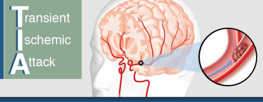
A transient ischemic attack (TIA) or commonly referred to as a mini-stroke has traditionally been regarded as a minor and temporary condition, but timely MRIs have proven that these events belong on the same spectrum as strokes. However, because TIAs, by definition, only last a short time, MR imaging must take place as quickly as possible for the fullest yield of useful information.


There is now a consensus that having a TIA increases a person's risk of a stroke.


Approximately one in six people who survive a TIA suffer a stroke within 90 days. Undergoing an MRI as soon as possible after a TIA can detect crucial warning signs that computed tomography (CT) alone cannot see.


Previous consensus guidelines from the American Heart Association (AHA) do not recommend MRI for all TIA patients because of the higher cost. However, mounting evidence suggests that an MRI within 1 to 2 days of a TIA could spot evidence of a stroke that may disappear in time.


MRIs can detect tissue damage even when symptoms are temporary.


The sophisticated imaging technique can detect stroke lesions that may become less apparent quickly. A study from the journal Stroke followed 263 patients who had suffered a TIA or minor stroke and received a baseline MRI within 24 hours. After 90 days, a follow-up MRI was conducted.


The results of each patient's two MRIs were assessed independently and the results confirm the importance of early scans. Thirty percent of patients with a negative scan at 90 days had a clearly identifiable stroke in the baseline image. Without the early scan, physicians would not know that a stroke had occurred in this large group of patients.


In spite of this evidence, some physicians settle for a less-precise CT scan. A recent study from Neurology found that just 40 percent of patients with TIA or minor stroke had an MRI performed within 48 hours.


New guidelines offer options for those at high-risk of stroke.


The AHA and American Stroke Association have published new consensus guidelines for preventing strokes in patients with a history of strokes or TIA. Reducing hypertension and statin therapy remain at the top of the list. Increasing physical activity, reducing sodium intake, and following a Mediterranean-style diet (as opposed to a low-fat diet) are recommended. Other practices, such as sleep assessments and anti-platelet therapy immediately following a TIA may be considered.


For patients with a history of stroke or TIA, the average annual rate of future stroke is at an all-time low. That's great news, but a more nuanced understanding of TIAs and timely MRI of those who suffer them could yield even more impressive results.


References:


AHA and ASA Release Guideline for Prevention of Future Stroke in Patients with Stroke or TIA. American Family Physician. January 2015;91(2):136-137. Available from: https://www.aafp.org/afp/2015/0115/p136.html


Chaturvedi S et al. Have clinicians adopted the use of brain MRI for patients with TIA and minor stroke? Neurology. January 2017;88(3):237-244. doi:10.1212/WNL.0000000000003503


Krieger D. Should patients with TIAs be hospitalized? Cleveland Clinic Journal of Medicine. August 2005;72(8):722-724. Available from: http://www.clevelandclinicmeded.com/medicalpubs/ccjm/august2005/krieger.htm


Moreau F et al. Early Magnetic Resonance Imaging in Transient Ischemic Attack and Minor Stroke: Do it or Lose it. Stroke. March 2013;44(3)671-674. doi:10.1161/STROKEAHA.111.680033