The Power of MRI Exams in Personal Injury Lawsuits


The health and wellbeing of individuals are often vulnerable to mishaps and accidents that can cause personal injuries. Such injuries could result in a legal dispute where the injured person, known as the plaintiff, seeks compensation for damages from the person or entity they believe caused their injury. In these instances, a comprehensive understanding of the injury's extent is paramount, often heavily relying on medical records and procedures. One such crucial diagnostic tool is the Magnetic Resonance Imaging (MRI) examination. This article delves into how MRI exams can provide substantial assistance in personal injury lawsuits.


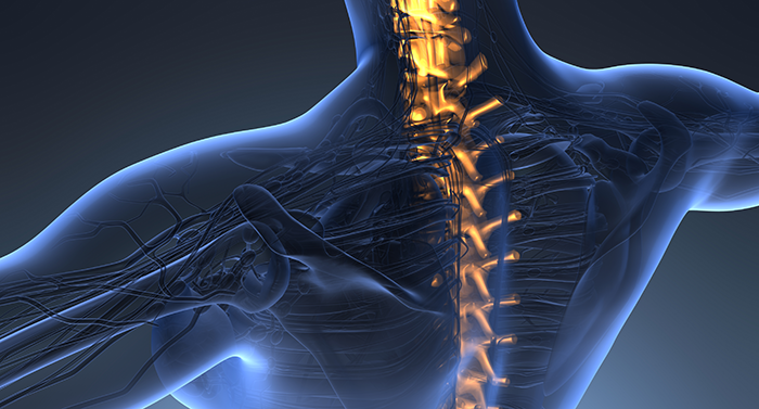
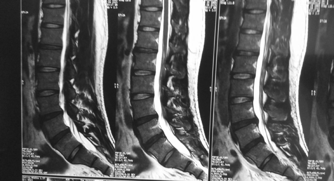
MRI is a non-invasive imaging technique that employs a powerful magnetic field and radio waves to generate detailed images of the inside of a body, typically focusing on soft tissues and organs. Unlike other imaging techniques like X-ray and CT scans, which primarily concentrate on skeletal injuries, MRI scans provide a comprehensive view of non-bony structures, like muscles, ligaments, tendons, and even the brain. This ability to visualize soft tissues makes MRI a valuable tool in evaluating the full extent of an injury.


In the context of personal injury lawsuits, MRI scans serve multiple critical roles. Primarily, it provides an objective basis for the plaintiff's claims of injury. Subjective symptoms like pain, discomfort, or restricted mobility may not convince a jury. However, visual evidence of damage, such as a torn ligament or herniated disc, becomes a tangible piece of evidence supporting the plaintiff's claims. By demonstrating the injury's existence and severity, MRI results can counter any argument that the plaintiff is exaggerating or feigning their condition.


Secondly, MRI results can contribute to determining the prognosis of an injury. The extent of damage revealed in the scan can help predict the duration of recovery, potential complications, and the possibility of permanent disability. This information is crucial in estimating the future medical costs, loss of earnings, and even non-economic damages like pain and suffering, which the plaintiff may include in their claim for compensation.


Furthermore, the timing of an MRI can also be a strategic element in a personal injury case. Early MRI exams might not reveal certain types of injuries like soft tissue damage, as these can take time to manifest. An MRI performed too soon could lead to an underestimation of the injury's extent, possibly harming the plaintiff's case. On the other hand, an MRI conducted too late might give the defendant an opportunity to argue that the injury was caused by something other than the initial incident. Hence, the timing of MRI is a critical factor in personal injury cases.


Finally, it's important to note that MRI results need to be interpreted and presented by a medical expert. Precise Imaging has several board certified radiologist that will review the images taken by the technologist and provide a report with their finding.


In conclusion, MRI examinations can be a powerful tool in personal injury lawsuits, aiding in confirming the existence and severity of an injury, predicting its prognosis, justifying medical expenses, and providing compelling evidence for the jury. However, these benefits are maximized when the timing of the MRI and interpretation of its results are strategically managed. It's crucial for individuals in these situations to consult with both medical and legal professionals to ensure the most effective use of MRI examinations in their case.


Diagnostic Imaging and Pre-Existing Conditions in Car Accident Claims


Diagnostic imaging is a powerful tool for car accident claims — but that’s true for insurance companies as well as plaintiffs. Personal injury attorneys often seek MRI scans and X-rays to demonstrate undeniable proof of injury. On the other hand, insurance companies will pounce on any hint of a pre-existing condition to avoid hefty payouts.


Don’t let insurers confuse the jury on the subject. In most states, a long-standing legal doctrine upholds the plaintiff’s right to damages in car accident cases. Pre-existing conditions are rarely the lone cause of injury for victims of car wrecks, and aggravating a condition still counts as injury.


Here’s what you need to know about car accident claims involving evidence of a pre-existing condition uncovered during imaging tests.


Degenerative Disc Disease in a Car Accident Case


Suppose you have a client who has been in a car accident, and is now suffering from lower back pain. An MRI scan reveals lumbar strain, but it also shows clear signs of degenerative disc disease. The insurer is likely to argue that the pre-existing condition reduces their liability for the injury.


This is by no means a rare scenario. Degenerative disc disease is extremely common; in fact, disc degeneration is all but inevitable for people older than 60. But many people who technically meet the diagnostic criteria for the condition don’t experience any symptoms at all.


The question under the law is not whether the pre-existing condition caused the injury; it is to what extent the accident produced the damage. The patient’s susceptibility to injury should not affect the value of the settlement; the court must consider the extent of the injury, not the plaintiff’s fragility.


The Eggshell Doctrine in U.S. Personal Injury Law


The Eggshell Doctrine is well established in most states. The maxim holds that defendants remain responsible for at-fault injuries regardless of the plaintiff’s pre-existing health conditions. In the above example, this would mean that, if the car accident led to a lumbar sprain in the victim, the presence of asymptomatic degenerative disc disease should not invalidate the claim.


In short, there’s no need to worry that an MRI scan, X-ray, or other form of diagnostic imaging will harm the plaintiff’s case. Even if advanced imaging techniques reveal pre-existing conditions, if the victim was injured in a car accident and another driver was at fault, the case remains strong.


Precise Imaging works with personal injury attorneys to obtain restitution for plaintiffs in multiple states. We offer the experience, flexibility, and patient-centered care it takes to win car accident cases, and we offer a suite of resources just for attorneys. Browse our online tools for lawyers here.   


To book an appointment for a client, call Precise Imaging at 800-558-2223 today.


Digital Sharing for Medical Images: PACS vs. VNAs

Digital Sharing for Medical Images: PACS vs. VNAs


Health care providers are virtually required to have digital infrastructure that includes sharable medical images these days. But if you’re looking to upgrade your system, or you’re implementing your first digital image archive, you have many software products to choose from.


The leading technologies for storing and retrieving medical imaging files are picture archiving and communications systems (PACS) and vendor neutral archives (VNAs). While the two competing formats look similar on first glance, there are notable differences between them. Before we get to the contrasts, though, it’s important to note what PACS and VNAs have in common. Here are a few of the constants across both types of imaging technologies:


  • Both PACS and VNAs provide remote access to images. Radiologists can upload images on one terminal, while physicians in a different office can log in to access them. This is the crucial requirement of all digital image-sharing systems.


  • Both systems operate with the same file format and transmission protocol: DICOM, which stands for “digital imaging and communications in medicine.” File formats can lead to accessibility problems, as each application is only equipped to handle certain types of files. The universal use of DICOMs between PACS and VNAs seems to suggest that migrating images between the two systems would be seamless. Unfortunately, that’s rarely the case. We’ll get into that a little later.


  • Both options may or may not also operate as a platform for non-DICOM images. This is an important question to ask when considering changes to an image-sharing system for medical care. If departments other than radiology are hoping to upload and access images through the same system, it is vital to choose software that can support non-DICOM images.


  • Designers of both PACS and VNAs are working to improve mobility and access to images across different types of devices. That’s one of the leading requests by radiologists and other health care providers — physicians want to be able to call up an image, safely and with full HIPAA compliance, on a tablet or even a phone in the examination room with their patients. They also want access at office desktops for consultation and reporting.


Despite these similarities, PACS and VNAs have very different sets of advantages. To complicate matters, many PACS are beginning to provide some of the features that initially launched the popularity of the VNA. Before making a system-wide purchase (or subscription service, which is also available from many vendors), it’s important to consult all stakeholders in your health care system, and to get to know your vendor and their product well.


That said, there are some broad-strokes difference between your average PACS and a typical VNA. We’ll get into those next.


PACS vs. VNAs: Differences Between Medical Image-Sharing Platforms


According to the trade publication Diagnostic Imaging, the VNA tends to focus on archiving and backing up data, while the PACS usually emphasizes workflow and user experience. Of course, these general principles are less significant in today’s market, where there are a wide variety of VNAs and PACS, and their strong points have begun to overlap.


That said, here are some of the differences between PACS and VNAs that users and medical industry analysts have pointed out:


  • The PACS is the original technology used to archive and retrieve digital medical images. As such, it’s often the choice of individual radiology departments, which were among the first to adopt digital imaging. VNAs, on the other hand, are more often found in multiple departments.


  • PAC systems are more highly proprietary than most VNAs. That is, each individual PACS will require its own user interface and its own log-in information. VNAs were designed to support storage and access across systems and vendors. Of course, new developments in PACS technology renders this difference conditional, but it remains the conventional wisdom among medical technology experts.


  • A VNA, by definition, divorces the storage/access functions of a PAC from a particular workstation or data silo. It uses its own application engine, allowing users to access images from multiple sources with the same user interface. That’s what makes VNAs typically better for interoperability between systems compared to a traditional PACS.


While the above list does seem to argue for the dominance of VNAs over PACS, in actual practice, implementation isn’t always the best choice for every provider. It can be expensive and time-consuming to migrate data from an existing PACS to a brand new VNA.


That’s because DICOM files contain both metadata and location pointers. The former attached patient information to the image; the latter helps the system find and pull up the specific image the user searches for.


During transition, all of this supplemental information can become scrambled, preventing access to images. In order to avoid this outcome, vendors often must reset DICOM headers and location pointers to ensure accuracy and access in the new system. That can be a lengthy and cost-intensive process.


Ultimately, then, institutions with the time and the money will benefit from an upgraded VNA system. More practically, some users will choose a PACS, with or without newly developed features. Regardless of the archiving and retrieval system, digital transmission for diagnostic images is practically a requirement in today’s medical system. Choose an imaging software product that makes it easy to share images with other departments and institutions.


References:


Jackson, Whitney. “What You Need to Know About VACS and VNA.DiagnosticImaging.” UBM, 4 Sept. 2014. Web. 15 Oct. 2018.  


O’Dowd, Elizabeth. “Pros and Cons of PACS, VNAs for Medical Image Data Storage.HitInfrastructure. Xtelligent Healthcare Media, LLC, n.d. Web. 15 Oct. 2018.


Will Artificial Intelligence Replace Radiologists One Day?

Will Artificial Intelligence Replace Radiologists One Day?


Artificial intelligence (AI) has undergone dramatic growth in recent years; its effects can be seen in virtually every field, from oil drilling to games of chess. Deep learning, a method which teaches computers to recognize objects within pictures, has significant potential impact on the medical imaging field.


That’s why some AI researchers say the technology could spell the end of human radiologists. Computers, they say, would be able to quickly, cheaply, and accurately perform the same job functions. While the power of AI cannot be denied, it seems premature to make assumptions about the important role trained human radiologists play in the contemporary health care industry. Artificial intelligence will almost certainly help radiologists do their jobs; we just doubt it can replace them entirely.


Radiologists may certainly be worried about the future of their profession, but any concern would be premature at this point. In fact, the adaptation of AI into imaging technology has great potential for increased productivity among radiologists and increased profitability for those institutions housing them. There are several current and future avenues in which AI may prove a useful augmentation to radiologists, as well as certain restrictions which ensure radiologists will continue to have gainful employment well into the future.


How AI Can Improve Radiology as the Tool of Human Radiologists


The first way in which AI may serve radiologists is in a quicker diagnosis of certain types of fractures and cancers. This would free the radiologist for more difficult-to-detect cases that could not be handled by current machine learning technology. In many cases, AI could also confirm a diagnosis that may have otherwise been in doubt, providing clinical decision support. The increased usage of deep learning in imaging also would allow the radiologist to spend more time focusing on the patient and their treatment plan, potentially creating greater job satisfaction for that individual.


A second major reason for the medical field to embrace AI is the potential for increased demand in imaging procedures. In countless cases throughout history, increased automation meant greater efficiency, which in turn brought the price of the given product down. Often throughout history, increased automation brought about fears of workers losing their jobs; however, the lower prices usually resulted in such a significant uptick in consumer demand that more jobs were created than lost. The same potential exists for the relationship between AI and radiologists.


Third, many conflate the type of AI learning being applied to radiology with the AI seen in science fiction movies. The deep learning being applied in to diagnose fractures or occurrences of cancer is an extremely focused technology used for that purpose and that purpose only. The AI is not suggesting a treatment pattern, or consoling a patient who has received devastating news. AI is only reading an object in a picture, albeit to a highly accurate degree in some cases.  


Why Human Radiologists Will Always Be Part of Diagnostic Imaging


No machine will ever be able to replace the empathy that medical providers develop throughout their careers. There’s no substitute for a warm hug, a meaningful conversation, or time spent with a trusted physician.


Artificial intelligence has the potential to greatly alter the field of radiology in many ways that generate a positive impact for all those involved, from the patient to the radiologist to the medical institution. Imaging specialists need not worry about job loss at the hands of a faceless algorithm. Instead, opportunities to increase patient well-being and diagnoses accuracy should be adopted whenever possible.      


How Diagnostic Imaging Could One Day Affect Tax Laws for Personal Injury Settlements

How Diagnostic Imaging Could One Day Affect Tax Laws for Personal Injury Settlements


With the 2017 passing of the Tax Cuts and Jobs Act, IRS tax policy on personal injury cases is back in the spotlight. At the same time, for the past few years, the legal profession has been absorbing the impact of ever-more detailed imaging studies that, some argue, display a physical basis for certain neurological phenomena, including disorders like PTSD and subjective experiences like pain itself.


Strangely, these two seemingly unrelated forces — tax laws for personal injury cases and the use of advanced imaging technologies in the courtroom — could combine to affect the take-home damages that plaintiffs receive from a successful lawsuit. Here's how, in a point-by-point analysis:


  • Tax laws for compensatory damages are complex, even circular. According to the IRS, damages paid out for physical injuries are tax-free. Damages for emotional injury, however, are fully taxable.


  • As researchers uncover more about the complex unity of physical and emotional systems in the human experience, courts and the IRS are faced with a challenge. Emotional trauma often leads to physical symptoms. Likewise, bodily injuries often result in emotional side effects. The two are difficult to separate. So which set of compensatory damages can legally go tax-free?  


  • Under current precedent, the IRS holds that taxability is associated with the principal injury. Cascading effects are lumped in with the root complaint. So if a defendant physically injures or sickens the plaintiff, compensatory damages awarded in the case are tax-free, even if some of those damages are paid out for emotional effects associated with the injury. Conversely, if the defendant's negligence leads to emotional harm, and that psychological trauma manifests in physical symptoms, damages are fully taxable, even if some portion is awarded in compensation for bodily sickness arising from emotional distress.  


  • The Tax Cuts and Jobs Act raises the stakes on this issue of taxation. Plaintiffs who hire contingent-fee attorneys are now responsible for paying taxes on 100 percent of case-related payments — including that portion that goes to pay the lawyer. Under previous law, attorneys and plaintiffs each assumed their share of the tax liability associated with taxable payments resulting from the same case.


  • Here's where advanced imaging could change the landscape. In 2008, the Arizona Superior Court in Pima County heard a landmark case, Koch v. Western Emulsions, Inc. The court allowed evidence of chronic neuropathic pain obtained with an fMRI scan, which the plaintiff's expert witness said showed a clear physical pattern of brain activity "proving" the existence of the pain. Ultimately, the defendant settled for $800,000, more than 10 times their initial offering.


  • Since Koch, the scientific community has warned against using advanced imaging as a sort of "painometer." The science just isn't there yet, and there are ethical issues involved in imposing "mind-reading" technologies on plaintiffs, researchers say.


  • But brain scans are showing physical changes associated with a lot more than pain. In one study, MRI scans showed changes in the physical structure of the hippocampus in  Vietnam veterans with PTSD. Some had an 8 percent reduction in volume of the right hippocampus.


The question this all leads us to, then, is where to draw the line between physical and psychological injury. Advanced imaging technologies will continue to show greater levels of detail in the troubled brain. At some point, the courts and the IRS will have to wade into one of the most philosophically murky questions of all time: the mind-body problem. More to the point: Is there any injury, psychological or otherwise, that cannot be described as physical?


The answers we come up with will have dramatic effects on the value of settlements, damages, and tax payments in the years to come.


5 Ways Physicians Can Keep Imaging Costs Down for Patients

According to the American Medical Association (AMA) Code of Medical Ethics, "Managing health care resources responsibly for the benefit of all patients is compatible with physicians' primary obligation to serve the interests of individual patients." In other words, doctors should consider the cost of treatment, and save their patients money when they can.


At a time of record health care costs — more than $28,000 for the typical U.S. family in 2018 — this isn't just an issue of saving patients a few dollars here and there. When patients can't afford to pay their health care bills, they're more likely to delay seeking treatment. That delay can affect the outcome of eventual care.


Of course, as the AMA states, "Physicians' primary ethical obligation is to promote the well-being of individual patients." Sometimes that well-being hinges on cash or its lack. So how can general practitioners and other referring physicians limit the cost of care for their patients? Diagnostic imaging is a great place to start. The cost of imaging studies has grown faster than wages, overall inflation, and health care expense.  


Luckily, doctors are pushing back against hospital pricing for these services. Here are a few ways referring physicians can provide excellent care for their patients without overspending on imaging studies:


  • Involve patients in the decision to seek or omit imaging tests. When a patient presents with conditions that aren't life threatening, doctors have significant leeway to work within the patient's preferences. Often, this leads to fewer imaging studies, with associated savings. In a recent study, doctors who used shared decision-making tools with their patients ordered 7 percent fewer advanced imaging tests and 30 percent fewer standard imaging studies.  


  • Avoid ordering full-body scans to screen for tumors unless patients show symptoms. The American College of Preventive Medicine discourages the use of whole-body scanning to screen asymptomatic patients for tumors. They point out that no data suggests survival improvement for patients, and that less than 2 percent of asymptomatic patients screened had tumors.


  • Choose imaging providers with upfront pricing, and share that information with patients. It shouldn't be difficult to find pricing for imaging procedures before making a referral. If a provider conceals prices, choose another imaging clinic. With accurate pricing information in hand, physicians can work with patients to conduct a cost-benefit analysis of ordering a study.


  • Refer patients to high-quality imaging clinics rather than relying on hospital radiology departments. As we've mentioned in this space before, an MRI from a freestanding independent imaging center is often thousands of dollars less than the same procedure at a hospital — even with the same doctors and the same equipment. Choosing a high-quality imaging provider that's free from hospital pricing is the easiest way to save patients money on diagnostic tests.  


  • Take advantage of digital delivery of diagnostic images and radiology reports. Doctors and patients can access digital reports anywhere and at any time, leading to lower costs and greater access. Precise Imaging offers doctors digital access and 24/7 tech support through a dedicated physician's portal.


With the ongoing public discussion of the U.S. health care system, awareness of the role of finances in treatment continues to rise. It's time to have these discussions; as of 2013, only 36 percent of surveyed physicians believed they had a "major responsibility" to control care costs on their patients' behalf.


But we know that affordability can translate into real-world effects on outcomes. Patient well-being and the costs of care are not two separate issues; they are bound together in complex, intractable ways. By choosing dedicated, patient-centered providers like Precise Imaging, doctors can get patients the services they need without unnecessarily adding to their burden of medical debt. Call us at 800-558-2223 or fill out our online form to make a referral today.   


References:


Bradley D, Bradley K. The value of diagnostic medical imaging. North Carolina Medical Journal [serial online]. March 2014;75(2):121-125. Available from: MEDLINE Complete, Ipswich, MA. Accessed August 16, 2018.


Choosing Wisely, an initiative of the ABIM Foundation. American College of Preventive Medicine: Five things physicians and patients should question. Choosing Wisely. [online]. February 25, 2015. Available from www.choosingwisely.org. Accessed August 16, 2018.


Herdman MT, Maude RJ, Chowdhury MS, et al. The Relationship between Poverty and Healthcare Seeking among Patients Hospitalized with Acute Febrile Illnesses in Chittagong, Bangladesh. Ali M, ed. PLoS ONE. 2016;11(4):e0152965. doi:10.1371/journal.pone.0152965. Accessed August 16, 2018.


O'Reilly K. The AMA Code of Medical Ethics and health care spending. AMA Wire. [serial online]. April 18, 2018. Available from wire.ama-assn.org. Accessed August 16, 2018.


Rainey M. Health care costs for typical American family hit record high. The Fiscal Times [serial online]. May 23, 2018. Available from www.thefiscaltimes.com. Accessed August 16, 2018.


Smith-Bindman R, Miglioretti DL, Larson EB. Rising Use Of Diagnostic Medical Imaging In A Large Integrated Health System: The use of imaging has skyrocketed in the past decade, but no one patient population or medical condition is responsible. Health affairs (Project Hope). 2008;27(6):1491-1502. doi:10.1377/hlthaff.27.6.1491. Accessed August 16, 2018.


Tilburt JC, Wynia MK, Sheeler RD, et al. Views of US Physicians About Controlling Health Care Costs. JAMA. 2013;310(4):380-388. doi:10.1001/jama.2013.8278.


The World Bank. Poverty and health. The World Bank. [online]. World Bank. August 25, 2014. Available from www.worldbank.com. Accessed August 16, 2018.


Veroff D, Marr A, Wennberg DE. Enhanced support for shared decision making reduced costs of care for patients with preference-sensitive conditions. Health affairs (Project Hope). 2013;32(2). doi.org/10.1377/hlthaff.2011.0941 Accessed August 16, 2018.


MRI Scans for Personal Injury Lawsuits: 4 Things to Look for in a Provider

Personal injury attorneys rely on MRI scans and other types of diagnostic imaging to build their cases. But not every hospital or clinic has the experience, the infrastructure, and the staff to provide crucial evidence when you need it.


Precise Imaging specializes in working with legal professionals while providing their clients with friendly, patient-centered service. We provide a single point-of-contact for management of all your personal injury cases, no matter how many you have at a time. We also have a designated web portal specifically for attorneys.


When you look for a diagnostic imaging center to work with, here are a few of the benefits you should look for — all of which you can find at Precise Imaging:    

1. Patient-Centered Care


Diagnostic imaging built around the patient's health and comfort is at the core of Precise Imaging practice. Our radiologists, technologists, and support staff are all trained to communicate warmly and with respect for the patient's values and experience. We take cues, both verbal and nonverbal, and offer emotional support alongside physical comforts to ensure that patients can get back to their busy lives feeling good about the experience.


Patient satisfaction is always our goal. We understand that attorneys are motivated by their clients' success because we feel the same.

2. Flexible and Convenient Scheduling


Speaking of patient satisfaction, market research suggests that patients list scheduling difficulty and waiting time for appointments as major issues in their choice of health care provider. We suspect that attorneys who make appointments for their clients would say much the same.  


With over 70 locations, Precise Imaging offers appointments for your clients when and where it's convenient for them. We can even schedule same-day imaging. Our radiologists provide reports within 24 hours of a scan — and faster when requested. Precise Imaging has years of experience working with the legal community, and we pride ourselves on taking the stress off patients and their representatives both through flexible scheduling.


When a client is struggling with an injury and a legal case, it helps to keep one thing, at least, as simple as possible. Our scheduling process is quick, easy, and convenient, no matter who makes the call.

3. HIPAA-Compliant Online Access to Images


Once a scan is completed and the radiologist analyzes the results, we offer images in the format of your choice. We can put the images on a CD or other digital device, or we can simply make them available to you, 24/7, through our attorney web portal.


The portal complies with all HIPAA regulations, allowing patients, health care professionals, and attorneys access to the information they need when they need it. Our attorney portal offers a user-friendly interface designed to simplify the organization of complex legal data.

4. Medical Lien Payments and Letters of Protection


Of course, unexpected medical bills can be difficult to cover. Your clients shouldn't worry about that while focusing on their cases. Precise Imaging accepts medical liens and letters of protection for personal injury to simplify patient finances during a trying time.


Precise Imaging offers a full suite of resources for attorneys. Find them here. And when you need MRI scans for personal injury lawsuits, contact Precise Imaging at 800-558-2223.


Understanding the Personal Injury Lien as Payment for Diagnostic Imaging

Personal injury liens help victims obtain medical treatment and stay afloat financially while their case is being decided. While this agreement entitles medical centers to repayment after a case has been settled, it can be greatly beneficial to attorneys and their clients as well.


Because many people cannot afford to pay major unforeseen medical bills, liens provide a lifeline until a settlement is reached, and attorneys benefit by ensuring that their clients get medical procedures and imaging completed in a timely manner. Still, there are a few things to consider when advising clients on this complicated subject.


The first thing to consider is what kind of experience the health care facility has with liens. Many smaller medical centers and imaging facilities don't accept personal injury liens; others may have a poor history of working out reimbursements. While the client may not have a choice of their initial point of care, they can certainly choose where subsequent imaging is done.


And that medical imaging is important for not only the person's future medical care, but also the outcome of their personal injury case.

Magnetic resonance imaging (MRI) scans can make or break a case.


Some physicians are reluctant to order MRI scans for accident victims — they feel that the scanning technology is so good at detecting problems that it can reveal false positives through asymptomatic abnormalities. However, there are many reasons that American courts encourage MRI scans.


For one, an MRI scan is completely objective. While radiologists may have differing interpretations of the scans, the scans themselves cannot be manipulated to distort the truth. That makes for powerful evidence to a judge or jury. Seeing the results of negligence in a black and white scan trumps many other forms of evidence.


Furthermore, seeing clear evidence of an injury can increase the value of a claim. Injuries in the brain, spinal discs, nerves, and joints can be difficult to verify without this state-of-the-art imagery. An MRI shows what many other forms of imaging cannot, and that's makes it invaluable to winning and maximizing personal injury lawsuits.

Of course, some patients may be reluctant to get MRI scans for a variety of reasons.


In these cases, victims of injury should be aware of how beneficial these scans can be for correct diagnosis and beginning recovery (not to mention, winning a case). Magnetic resonance allows doctors to pinpoint the source of an injury with the very latest technology available.


Some people hesitate to complete scans because of anxiety over claustrophobia. These fears are completely understandable, but there has been progress in recent years in making the technology more patient-friendly. For instance, open MRIs can help keep patients calm by eliminating the closed spaces that exist in traditional MRI machines. Even simple strategies, like prescribing an anti-anxiety medication or providing headphones with music can help anxious patients get through a scan.

Choosing an imaging center with personal injury experience can make the process easier for patients and attorneys.


The biggest reason is that facilities with experience offer a patient-centered focus and streamlined approach to accessing medical images. These facilities understand HIPAA regulations, know the preferred formats for images, and offer web portals for easy accessibility.


Using a single, preferred diagnostic company can also simplify caseloads for attorneys.

By relying on a trusted imaging center, firms know that crucial evidence is always just a click away — no matter what time of day. Attorneys can also trust that their clients are getting the best rates, so that injury victims can hold onto the lion's share of their settlement.


Finding a diagnostic imaging center that can handle time-sensitive requests is also important. Many hospitals experience lengthy delays in scheduling scans. Because Precise Imaging has over 70 locations, we can schedule same-day scans. Even better, our radiologists will file reports no later than 48 hours after the scan (and sometimes much sooner). The quick scheduling and prompt reading allows personal injury cases to clear significant hurdles without taking up too much time.

Look for an imaging center with web portals for attorneys.


Because legal cases are time-sensitive, having a 24-7 web portal available is crucial. Precise Imaging provides such portals so that attorneys can access important information night or day. Our web portals provide medical images, payment information, and case details, all while fully complying with HIPAA regulations. This service keeps attorneys organized and on track without having to rely on the medical center being open for service.  


Our facilities are well-acquainted with the needs of personal injury lawyers, and our patient-centered facilities can comfort distraught patients who have recently suffered injuries. We offer a number of anxiety-reducing strategies to make our MRI scans as comfortable as possible.


We can schedule scans for the same day and get you results within 48 hours. All of the information an attorney or patient needs to access can be found on our 24-hour-per-day web portal. These services simplify a personal injury case and keep you on track.


We have partnered with patients and attorneys for thousands of personal injury cases and take our responsibilities to all parties seriously. We are prepared to accept all forms of payment, including workers' comp, liens, and deferred payment (through a letter of protection).


Precise Imaging makes scans fast and easy for patients and provides streamlined results for attorneys. If you'd like to partner with Precise Imaging for your personal injury cases, call 800.558.2223 or make an online referral here.


References


"7 Steps to Approaching Lien Claims in Personal Injury Cases." FindLaw.com. n.d. Web. 3 May 2018.


Coye Law Firm. "What Is A Letter Of Protection? Learn More About The Pros And Cons Of Using A Letter Of Protection Or LOP." HG.org n.d. Web. 3 May 2018.


Jacobson, Martin & Schweers, R. "Ethical Considerations in Personal Injury Settlements and Lien Resolution." Bill of Particulars. Vol. 1 2012. Web. 3 May 2018.


"Medical Liens: A Primer." The Steve Dhillon Law Firm. n.d. Web. 3 May 2018.


Weiner, Ron "Lien vs. Subrogation." MedLien Solutions. 3 Nov. 2015. Web. 3 May 2018.


Choosing Diagnostic Imaging Services for Personal Injury Cases

Magnetic resonance imaging (MRI) scans can be crucial pieces of evidence in personal injury lawsuits. The detailed images can convince a jury or judge that an injury occurred as a result of an accident or negligence, rather than aging and genetics.


MRI scans are universally accepted by insurance companies and courts because of their objectivity and superiority to older forms of imaging, like X-ray. Different radiologists may have differing interpretations of the scans, but in general, the results of an MRI scan provide definitive evidence to a judge or jury.


Another great advantage of MRI scans is that they use no radiation, and thus are safer than alternative forms of medical imaging. Instead, MRI scans use powerful magnets to develop highly detailed scans of the body. These powerful magnets require special attention for those with pacemakers or other implanted devices, but scans can still be safely given to most cardiac patients.


Litigation MRI scans are helping everyone from injured motorists to football players with traumatic brain injuries. It's this versatility that makes MRI scans the gold standard for determining medical history.

Unfortunately, MRI scanners at hospitals are expensive and in high-demand.


This may lead the doctor to prescribe rest for your client's injury before signing off on an MRI scan. If you'd like to move your case along, we can help schedule an affordable and timely scan. Precise Imaging has years of experience and accepts cases on lien. Advantages of working with us include:


  1. Fast and easy scheduling - Call Precise Imaging at (800) 558-2223 or schedule an appointment online. We have over 70 locations, many open nights and weekends, to get your scans done quickly and professionally.

  2. A variety of delivery options - Obtain scans on compact discs or other secure media for reliable access. We're experienced in handling potential evidence and same-day reporting is available in many locations.

  3. A range of imaging modalities - Whether you need dual-focus X-ray tubes or an open MRI machine, we've got you covered. For any type of medical imaging, we'll harness our flexibility to get you the best images for your case.

  4. Our facilities are patient-focused - We know that patients who have suffered an injury are under stress and possibly in pain. Our highly trained techs will make sure patients are comfortable and stress-free as they undergo imaging.


Precise Imaging has served more than 150,000 patients throughout California, Nevada, and Arizona. Whether you're an attorney or a patient, we can schedule an appointment at a convenient time and place. Call (800) 558-2223 or make an appointment online.


References


Cherniak, Todd. "Litigation MRI: Why Lawyers Are Asking for It and Why Your Patients Need It." British Columbia Medical Journal. Vol. 47, No. 7. Sept. 2005. Pp 358-361.


"Magnetic Resonance Imaging (MRI) Safety." RadiologyInfo.org. 5 Apr. 2017. Web. 1 May 2018.


"MRI is Safe for Most People with Pacemakers and Defibrillators." Harvard Health Publishing. Harvard Medical School. Jan. 2018. Web. 1 May 2018.


Brown, Francis H. "Recent Developments in Traumatic Brain Injury Litigation." American Bar Association. 12 Sept. 2016. Web. 1 May 2018.

MRI Technique Shows Brain Differences in People on Autism Spectrum

Researchers have observed abnormal neural networks in preschoolers with autism spectrum disorder (ASD) using a specialized magnetic resonance imaging (MRI) technique. The discovery gives hope that MRI scanning may one day allow early diagnosis, intervention, and treatment in ASD.

The study included 21 children with ASD and 21 children with typical development (TD).


It took place over four years in the Chinese PLA Hospital in Beijing. The researchers used a special MRI technique called diffusion-tensor imaging (DTI), which allowed them to observe the location, orientation, and anisotropy of white matter tracts in the children's brains.


Other scientists have used a similar technique to study the brains of patients with Alzheimer disease, multiple sclerosis, epilepsy, and other psychiatric disorders. The approach has led to

an improved understanding of topological organization of the brain network in patients with these disorders.


There were similarly useful results in the study of preschoolers with ASD. The researchers observed alterations of white matter in the children with ASD compared to children with TD. The scientists were able to correlate the alterations in the white matter networks with delays in verbal communication, object use, visual response, body use, and listening response.

The study also observed increased nodal efficiency in children with ASD compared with TD.


That observation agreed with previous studies that had observed this phenomenon in adults on the autism spectrum. The researchers believe this is a reflection of a delayed maturation process in people with ASD.


"Altered brain connectivity may be a key pathophysiological feature of ASD," study co-author Lin Ma told Science Daily. "This altered connectivity is visualized in our findings, thus providing a further step in understanding ASD. The imaging finding of those 'targets' may be a clue for future diagnosis and even for therapeutic intervention in preschool children with ASD."

Medical professionals from around the world weighed in on the study.


"This discovery gives us a more objective diagnostic method by using MRIs to aid us in the diagnosis of children who do have autism, and also gives us a better understanding of the abnormal differences in the brain," psychiatrist Dr. Matthew Lorber told HealthDay Reporter.


Doctors presently diagnose ASD through observing difficulties with social use of communication and interaction in children. A standardized MRI technique that could identify the disorder could give parents and therapists an earlier chance at intervention and treatment.  


It's still too early to rely on MRI scans for the diagnosis of autism, but the study's results show that it's possible. The study also provides other researchers with imaging biomarkers that may speed the development of this technology.


References

"Abnormal Brain Connections Seen in Preschoolers with Autism." Science Daily. 27 Mar. 2018. Accessed 29 Mar. 2018.


"DSM-5 Diagnostic Criteria." Autism Speaks. 2018. Accessed 29 Mar. 2018.


Ma, Lin et al. "Alterations of White Matter Connectivity in Preschool Children with Autism Spectrum Disorder." Radiology. 27 Mar. 2018. doi: 10.1148/radiol.2018170059. [Epub ahead of print]. Accessed 29 Mar. 2018.


Preidt, Robert. "MRI Sheds New Light on Brain Networks Tied to Autism." HealthDay Reporter. 27 Mar. 2018. Accessed 29 Mar. 2018.