mri-biopsy

Precise Blog – How MRI Scans Can Reduce the Need for Biopsies

How MRI Scans Can Reduce the Need for Biopsies


Researchers at the Simmons Comprehensive Cancer Center have authored a study detailing a multiparametric magnetic resonance imaging (mpMRI) technique that predicts a malignant type of kidney cancer without performing a biopsy. The results of the method are impressive, but require more refinement to fully take the place of a biopsy.


Doctors frequently find kidney tumors accidentally while conducting CT scans for other reasons.

These scans alone do not yield the necessary information that tells doctors whether they are malignant or benign. Instead, a biopsy is usually performed. These procedures can save lives by correctly identifying the nature of the tumor, but they are also invasive and can cause complications.


“Using mpMRI, multiple types of images can be obtained from the renal mass and each one tells us something about the tissue,” Dr. Ivan Pedrosa, Professor of Radiology and Chief of Magnetic Resonance Imaging told the UT Southwestern Newsroom.


Identifying malignant masses in the kidney is extremely important because treatment is highly effective before the tumor metastasizes. However, once it spreads to other parts of the body, survival rates are low. Clear cell kidney carcinoma is an aggressive subtype of malignant masses that the researchers.


Seven radiologists studied the records of 110 patients with cT1a masses.


These patients had all undergone an MRI as well as a partial or radical nephrectomy. The observing radiologists did not know the final pathology findings, but instead relied on an algorithm to judge whether tumors were metastatic.


The researchers had 78 percent accuracy when rating that the mass was "probably" or "definitely" clear cell kidney carcinoma. When rating that the mass was possibly carcinoma, they had a 95 percent success rate.


The promising results show that biopsies may not be necessary for identifying certain cancers.


Because some patients are reluctant to consent to biopsies, this new technique is potentially lifesaving. As it stands, patients who do not want a biopsy may learn important information if an MRI shows that their kidney mass has a high probability of becoming metastatic. This new information could convince them that the pain of a biopsy is worth going through.


Using these methods to identify clear cell histology is still a work in progress. The doctors at Simmons Comprehensive Cancer Center will have to achieve a higher degree of accuracy in predicting malignant kidney masses for the method to become mainstream.


However, as standardization of imaging protocols and reporting criteria are refined, accurate results should increase. When MRI scans alone are sufficient to identify clear cell kidney carcinoma, doctors will have another powerful tool in the fight against cancer.


References:


Anderson, Avery. “State-of-the-Art MRI Technology Bypasses Need for Biopsy.” UT Southwestern Medical Center, 2 Jan. 2018. Accessed March 14, 2018.

Canvasser, N. et al. "Diagnostic Accuracy of Multiparametric Magnetic Resonance Imaging to Identify Clear Cell Renal Cell Carcinoma in cT1a Renal Masses." The Journal of Urology, volume 198, no. 4, Oct. 2017, pp. 780-786. Accessed March 14, 2018.

National Cancer Institute. “Kidney Cancer.” Kidney (Renal Cell) Cancer—Health Professional Version. Accessed March 14, 2018.


National Institute of Health. “Clear Cell Kidney Carcinoma.” The Cancer Genome Atlas, National Institute of Health. Accessed March 14, 2018.

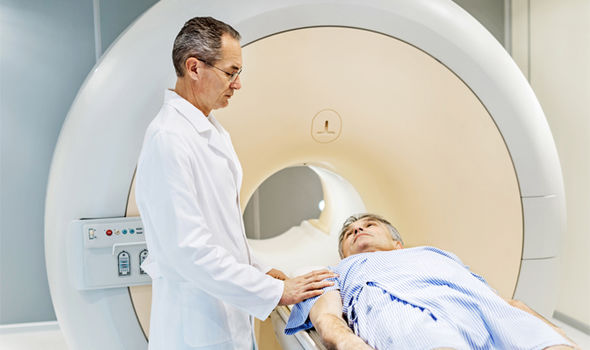
The MRI Procedure in 10 Steps: Managing Patient Anxiety Through Information


A first-time MRI procedure can make patients nervous, even to the point of ending the scan. That can lead to higher costs for imaging centers, and even affect patient outcomes if the anxiety interferes with the quality of the radiology report. Research shows one simple way to help nervous patients get through their scans without interruption: Communication.


A 2015 study in the journal Magnetic Resonance Imaging tested an intervention in which imaging staff explained the MRI process to one group of patients. They took blood samples during the scans, later testing them for the stress hormones prolactin and cortisol. Additionally, they took both the experimental and control groups through the 40-questions State-Trait Anxiety Inventory to measure patient nervousness.


The patients who had the intervention in which staff verbally shared information about the scan showed a 6-percent drop in cortisol after the scan. The control group's cortisol levels increased by 18 percent. The authors of the study conclude that "MRI anxiety can be reduced by information and communication. This combined method is shown to be effective and should be used during daily radiology routine."


So what can physicians do to prepare their patients for a first MRI scan in advance? It's never too early to start educating patients about what they can expect during a health procedure. And the MRI process can be boiled down into 10, easy-to-grasp steps. Share these steps with patients to help limit anxiety during an MRI scan:


  1. First, radiology staff will walk the patient through a detailed screening process. Because of the strong magnetic field generated during an MRI, patients must report any medical implants or metal particles in their bodies. These may preclude the use of MRI imaging. 

  2. Once the patient clears the screening, staff will lead them into the MRI suite. Some imaging facilities offer hospital robes, to ensure there's no metal in the patient's clothing. Others allow patients to wear their own metal-free clothes, such as sweat pants and a T-shirt. The technologist will proceed to position the patient on the table; most commonly, patients lie on their backs. If the scan requires an additional radiofrequency coil, the technologist will place that on the patient's body at this time.


  3. The patient enters the bore. Meanwhile, technologists cycle through a list of pre-programmed settings called "protocols." They'll choose the protocol that corresponds with the body part they are imaging; this will tell the MRI machine which angles, targets, and pulse sequences to use in this particular procedure.


  4. Before the scan proper begins, technologists run a "scout" or "localizer" scan. This is a low-quality image, and it won't be used in reporting. However, localizer scans obtain visual and placement information that the computer will used to plan the angles of its imaging later in the process.


  5. Parallel imaging is a process designed to speed up scan time. It collects less raw data during the scan, and patches missing information using special algorithms to generate the final image. Parallel imaging requires specifically calibrated coils, and may call for a calibration scan at this point.


  6. One of the great strengths of MRI scans is that they create 3D images that can be viewed from any angle. The next step is to program in the angle of images for the radiologist. Technologists can change the "thickness" of the image at this point, as well.


  7. Before the scanner can begin collecting valuable images, it must calibrate all systems through the use of a prescan. This shouldn't take much more than 10 or 20 seconds.


  8. It is only at this relatively late stage in the process that the technologist actually runs the scan. They will make necessary adjustments and continue scanning according to the chosen protocol. In the end, they'll have clear, accurate images that radiologists will use in their reporting.


  9. Some types of images require extra work in post-production, but this can be done after the patient has left the MRI suite.


  10. Scanning complete, the technologist pulls the patient from the bore. Different types of scans take varying lengths of time, but most range between 20 and 60 minutes.      


When patients understand more about their medical procedures, and know what to expect, they're less likely to experience significant anxiety. That's both a value in itself — as patient-centered caregivers, staff at Precise Imaging works to keep patients comfortable, both physically and emotionally — and an element of better diagnoses, which lead to better patient outcomes.


To learn more about an MRI procedure from Precise Imaging, or to refer a patient, call us at 800-558-2223.  


References:


Deshmane A, Gulani V, Griswold MA, Seiberlich N. Parallel MR imaging. Journal of Magnetic Resonance Imaging: JMRI. 2012;36(1):55-72. doi:10.1002/jmri.23639


Elster AD. "Performing an MR Scan." MRIQuestions.com. 2017. Web. Jan. 8 2018.


Tazegul G, Etcioglu E, Yildiz F, Tuney D. Can MRI related patient anxiety be prevented? Magnetic Resonance Imaging. 2015;33(1):180-3. doi:10.1016/j.mri.2014.08.024


Should Physicians Order MRI Scans for Personal Injury Litigation?

Should Physicians Order MRI Scans for Personal Injury Litigation?


Physicians are rightfully reluctant to order MRI scans on patients they can confidently diagnose using other means. The clarity of the MRI image is, paradoxically, the reason why many physicians use them sparingly; MRI scans reveal asymptomatic abnormalities that can lead to false positives if doctors are overzealous in their diagnosis.


However, there are compelling reasons why physicians should still bow to attorney requests for MRI scans in personal injury cases. In fact, according to a 2005 argument published in the British Columbia Medical Journal, MRI scans were remarkably common in U.S. personal injury cases. This is no time for that to change. Here's why:


  1. False positives can be controlled by the reporting radiologist's scrutiny and the health care team's other diagnostic efforts.  


MRI scans alone might result in inaccuracies, but physicians don't see these results in a vacuum. Medically, the MRI scan is one part of a larger diagnostic whole. Meanwhile, legally, the scan images can convince the court of the claim's validity.   


  1. MRI scans can provide clear, convincing evidence of injury in many cases.


There's no clearer way to show soft tissue damage than an MRI image, which can be colored to highlight areas of interest, and even displayed in three dimensions.


  • The use of MRI protects physicians from later claims of negligent care in the unfortunate case that a more serious injury is revealed in the future.


If an MRI scan could have revealed an injury, and the physician fails to order one, that could leave the doctor vulnerable to legal action.


  1. It's easy to schedule quick, accurate, and affordable MRI scans.


Legal cases happen on a strict schedule, which is why Precise Imaging ensures a quick turnaround on reports, same-day scheduling, and fair pricing.   


  1. The costs of MRI are often recoverable in personal injury cases.


All Precise Imaging locations accept payments via personal injury lien and letters of protection, so if the patient wins the case, the procedure is covered.   


The Value of MRI Images in Personal Injury Cases


MRI technology is a crucial tool for diagnosing problems with the brain, spinal discs, nerves, joints, and muscles that may not be detected using other methods. These soft tissues are often associated with personal injury cases stemming from car accidents or slips and falls. Once the medical team makes the diagnosis, recovery can begin — and the case can play out.  


From a legal perspective, the images produced by an MRI scan can help win cases and might even increase the value of the claim, which ultimately benefits patients, doctors, and attorneys. MRI results are objective and can be used as a helpful “final word” in a legal setting. By using MRI scans in mediation or at trial, lawyers can fight for what patients need to cover their health care, now and in the future. That's a worthwhile goal for physicians as well.  


To learn more, or to make a referral, call Precise Imaging at 800-558-2223.

5 Ways Radiology Technologists Reduce Patient Claustrophobia During MRI Scans

5 Ways Radiology Technologists Reduce Patient Claustrophobia During MRI Scans


Physicians strive to prevent distress in patients at every turn, but between 4 and 30 percent of patients who undergo MRI scans still report some level of anxiety. This isn't just a problem for patients; claustrophobic reactions can add considerable time to MRI scans, and can even adversely affect clinical findings through lower-quality images.


Even worse, according to a 2007 article published in the Journal of Magnetic Resonance Imaging, claustrophobic patients abort around 2 million MRI scans around the world every year. That deprives physicians of much-needed diagnostic images.  


The good news is that radiologists and technologists have developed interventions that can help to reduce claustrophobic reactions leading up to and during the procedure. As the first point of contact with patients, referring physicians can help lay the groundwork for a comfortable, anxiety-free patient experience by explaining how technologists can help alleviate their fears as soon as a patient expresses trepidation about the upcoming imaging exam.


To that effect, here are some of the latest techniques and strategies physicians can explain to nervous patients when they refer them for an MRI scan:


  1. The first step to treating anxiety is to recognize that it is occurring, and technologists are trained to look for signs of discomfort throughout the entire patient interaction.


A 2013 study published in the International Journal of Behavioral Medicine used both subjective self-reports before and after the procedure to gauge patient anxiety. The researchers also measured electro-physiological reactions during the entirety of the study scan. Their findings suggest that patients tend to be most anxious at the beginning of the scan, as they enter the MRI bore. Then their distress usually diminishes throughout the course of the procedure.


Physicians can explain that patients might feel nervous or unable to complete the MRI scan at the beginning, but that once they're through that portion of the scan, they'll likely begin to feel more calm.


However, patients should know that technologists are aware of the patient's experience, and are committed to keeping them comfortable. Imaging staff listen for verbal cues of patient anxiety — from an explicit admission of fear to a tremor in the voice — and will move quickly to help a patient through a moment of panic.


Technologists also watch their patients' eyes; people with claustrophobia might glance nervously at the scanner's bore or else avoid looking at the machinery altogether. Other indications of anxiety that technologists watch for include blanching or flushing in the patient's face; reluctance to follow simple instructions; sweating; or reports of tachycardia.


Physicians can help to prepare nervous patients for a better experience at the imaging center by assuring them that the imaging staff is aware of, and responsive to, any discomfort they might feel, and that they will pause the procedure at the patient's request.  


  1. Patients should know that the choice to go through with an MRI scan is entirely within their power; technologists will never try to coerce them to go through with a procedure they decide to delay.


Ultimately, health care choices belong to the patient. If patients express anxiety about an upcoming MRI scan, physicians can assure them of this fact. They can explain that the patient can choose to forego a scan at any point during the treatment process.


By including this comforting information in an explanation of the health benefits of the MR images, physicians both place the nexus of control within the patient while also gently encouraging them to undergo the procedure for the sake of their health.   


  1. Technologists provide detailed, thorough explanations to patients before entering the examination room.


Patients who know what to expect are less likely to become anxious. But technologists also use their pre-scan conversations to build healthy clinical relationships with patients. It's important for patients to know that their technologists are friendly, understanding, competent, and utterly trustworthy. Early conversations help to create this welcoming atmosphere.


Physicians can lay the groundwork for a successful imaging procedure by assuring their patients of the technologist's positive attributes. That's why it's so valuable for physicians to work with preferred imaging providers, building relationships and trust that they use to help reassure patients who might be uncomfortable with the procedure. It's all part of the patient-centered approach that Precise Imaging practices.


  1. There are multiple distraction-based interventions that have documented history of helping patients through their anxiety when they enter the scanner bore.


The authors of Pinpointing Moments of High Anxiety During an MRI Examination, the study from the International Journal of Behavioral Medicine, seem hopeful about in-bore audiovisual systems that could distract patients during moments of anxiety — perhaps by allowing them to experience favorite TV shows or movies during the exam. The researchers also point to the use of prism glasses, which provide mirrors that allow patients to see outside of the bore, even as they lay flat for the procedure.


However, even something as simple as placing a soft cloth over the patient's eyes can help alleviate claustrophobia, writes technologist Thomas Rotunda, BSMI, BSHA, R.T.(R)(MR)(QM), in a recent edition of the journal Radiologic Technology.


This practice "is a basic way to combat claustrophobia because it helps patients ignore how close the inside of the scanner is to their faces, which likely makes them feel less confined," Rotunda writes.


  1. Newer scanning technology can be incredibly effective in reducing claustrophobia among patients.


One of the most powerful changes that imaging centers can make is to update their equipment, and that's been true for a decade. A seminal 2007 study in the Journal of Magnetic Resonance Imaging found that the latest MRI scanners, which reduced noise in-bore noise by 97 percent while also featuring a shorter bore than previous models, could reduce the incidence rate of claustrophobia by three times.


Physicians can reduce the expectation of anxiety in their patients by explaining the changes that have taken place since the old days of narrow bores and loud machines. Managing patient expectation will go a long way toward reducing claustrophobia and anxiety, which will lead to greater outcomes for patients.


Call Precise Imaging at 800-558-2223 to discuss advanced imaging equipment available and anxiety-reduction options, or to make a referral.


References:


Dewey M, Schink T, Dewey C. Claustrophobia During Magnetic Resonance Imaging: Cohort Study in Over 55,000 Patients. Journal of Magnetic Resonance Imaging. November 2007; 26(5):1322-1327. Available from: Wiley Online Library. Accessed November 2, 2017.


Melendez J, McCrank E. Anxiety-related reactions associated with magnetic resonance imaging examinations. JAMA, The Journal Of The American Medical Association [serial online]. 1993;(6):745. Available from: General OneFile, Ipswich, MA. Accessed November 2, 2017.


Minde D, Klaming L, Weda H. Pinpointing Moments of High Anxiety During an MRI Examination. International Journal Of Behavioral Medicine [serial online]. June 2014;21(3):487-495. Available from: CINAHL Plus with Full Text, Ipswich, MA. Accessed November 2, 2017.


Rotunda T. Reducing Occurrences of MR-related Claustrophobia in Patients With PTSD. Radiologic Technology [serial online]. September 2017;89(1):97-99. Available from: CINAHL Plus with Full Text, Ipswich, MA. Accessed November 2, 2017.

 
No health Insurance

Diagnostic Imaging Referrals for Uninsured Patients

What should physicians do when they suspect a patient needs an expensive diagnostic imaging procedure, but they also know that patient lacks health insurance, or has an inadequate plan? How do they weigh financial considerations against crucial diagnostic information?


As physicians, you balance a huge number of variables in deciding which diagnostic imaging procedures to order for your patients. You must weigh radiation exposure versus probability of actionable images, for instance, or decide whether a patient needs an X-ray, an MRI, or both.


And like it or not, every decision a referring physician makes will have an impact on the patient's final bill — which becomes problematic, both ethically and medically, given the role financial stress plays in negative health outcomes such as depression.


Lack of Health Insurance and Frequency of Diagnostic Imaging


Here's what we do know, at least as of the 2012 publication of this study from the Journal of the American College of Radiology: Uninsured Americans received fewer imaging procedures in emergency departments than insured patients. In a sense, this is unsurprising; Americans without health insurance generally receive less health care overall.


What we don't know is what to do about it. Should we order less imaging for insured patients, or more for uninsured patients? We won't really know until we can measure the outcomes of uninsured patients who receive equal care. To do that, we must find a way to provide truly affordable diagnostic imaging for all patients. That's where Precise Imaging can help.  


How Physicians Can Reduce the Financial Impact of Imaging on Patients


The ideal situation would be for physicians to have access to imaging centers that offer a full and flexible range of payment options along with the highest-quality service, simple scheduling, and fast results. That's exactly the combination that Precise Imaging offers to referring physicians.


Precise Imaging operates 70+ locations with evening and weekend hours, so your patients can always find a time and a place that suits them. If transportation is a problem, Precise Imaging can send a car for free. And with same-day scheduling and a 24-hour average turnaround on reports, you'll have the information you need without delay.


But the real issue for uninsured patients is the financial burden. That's why Precise Imaging's experienced billing team is trained and authorized to handle an incredible variety of payment options. For personal injury cases, they can accept liens. If workers' compensation is involved, that's no problem.


Precise Imaging even offers special cash prices for uninsured or underinsured patients. Friendly billing staff can also work with payors to establish structured payment plans. These options can create powerful discounts for patients in need, and might be just the assurance that you need to order a procedure you're on the fence about.


Financial considerations shouldn't have any place in patient care, but, unfortunately, they are real, and must be considered. Next time you're weighing the options for an uninsured patient, remember that Precise Imaging can help. Call Precise Imaging at 800-558-2223 to learn more about cash prices for uninsured patients.    


References:


Moser, James and Kimberly Applegate. "Imaging and Insurance: Do the Uninsured Get Less Imaging in Emergency Departments?" JACR. Journal of the American College of Radiology, Jan. 2012. Web. 29 Sept. 2017.


Galea, Sandro et. al. "Urban Neighborhood Poverty and the Incidence of Depression in a Population-Based Cohort Study." ScienceDirect. Annals of Epidemiology, Mar. 2007. Web. 29 Sept. 2017.

 
Online Portal

Why Online Radiology Portals are Good for Physicians and Their Patients

Online radiology portals are slowly moving into the mainstream. The advantages of these information-sharing platforms are clear. Physician portals allow quick, simple referrals and real-time access to diagnostic images, plus immediate interaction for remote members of health care teams. Patient portals give recipients of diagnostic imaging procedures unprecedented information, control over their own health care decisions, and more direct interaction with radiologists.


But are online health portals really working for patients and their physicians? What does the science say about the benefits and/or drawbacks of these tools? A recent surge of studies gives us some idea. Here are a few takeaways from the latest research on radiology portals:


  • An early study, published in the Journal of the American College of Radiology in 2015, concluded that "Referring-physician release of radiology reports via the online portal is important to patients, useful to referring physicians, and does not affect referring-physician workloads." However, the study noted, physicians may wish to delay the release of the radiology reports until after they've had the chance to discuss the findings with the patient.


  • A more recent study from the American Journal Of Roentgenology suggests that online patient portals create more communication between radiologists and patients themselves. This could be a profound shift; traditionally, radiologists confer with physicians, who then speak face-to-face with patients. Through the use of online radiology portals, patients may benefit from more direct interaction with their radiologists. "Radiologist collaboration with referring physicians is important in providing care in accordance with patient preferences," the study cautions.


  • A 2016 article from the Journal of the American College of Radiology picks up this theme of the changing role of the radiologist in an era of online portals. When patients have direct access to radiology reports, they'll likely have questions and/or concerns.


"Ultimately, radiologists may have to stop thinking about the radiology report as their final product and, instead, start thinking about the report as a springboard for becoming more active health care partners," concludes Christoph Lee et. al., authors of Implications of Direct Patient Online Access to Radiology Reports Through Patient Web Portals.


These changes provide benefits to patients and referring physicians alike, the authors suggest: "The published evidence to date suggests that the potential benefits likely outweigh the risks of providing patients with direct online access to their medical records, including their radiology reports."


Online Radiology Portals for Physicians and Patients at Precise Imaging


Every Precise Imaging location offers physicians access to a robust online portal. There, doctors can make quick referrals, receive radiology reports, and interact with the radiologists who write those reports. We offer 24/7 IT support and simplified payment modalities.


Meanwhile, patients can track their own health care journey through our fully HIPAA-compliant patient web portal. With these digital tools from Precise Imaging, patients, physicians, and radiologists all remain on the same page, striving for better outcomes.


Learn more about Precise Imaging's online portals by calling 800-558-2223 today.


References:


Gefen, Ron, Michael bruno, and Hani Abujudeh. "Online Portals: Gateway to Patient-Centered Radiology." AJROnline. American Journal of Roentgenology, July 2017. Web. 18 Sept. 2017.   


Henshaw, D., et. al. "Access to Radiology Reports via an Online Patient Portal: Experiences of Referring Physicians and Patients." PubMed. Journal of the American College of Radiology, June 2015. Web. 18 Sept. 2017.


Lee, Christoph, et. al. "Implications of Direct Patient Online Access to Radiology Reports Through Patient Web Portals." JACR. Journal of the American College of Radiology, Dec. 2016. PDF. 18 Sept. 2017.

The Benefits of Large Imaging Networks vs. Small Independent Facilities

Precise Imaging operates more than 70 locations in California, Arizona, and Nevada. The size of this network provides benefits to referring doctors and patients alike.


Smaller, independent imaging facilities often can't produce the same advantages — they simply don't have the staff, the space, or the infrastructure. The advantages of a large-scale diagnostic imaging organization like Precise Imaging include:

 
More convenient scheduling. 


Precise Imaging locations are open late and on weekends, and they're distributed all over major metropolitan areas. This makes it easier and more convenient for patients to schedule an imaging appointment close to them, and at a time they prefer. Referring physicians can even ask for same-day service for cases in which time is of the essence.

 
Simplified billing.


Medical billing is complex enough. Rather than filing paperwork with many different small-time providers, choosing Precise Imaging gives medical providers a single contact point for all issues of billing. Precise Imaging offers a clear and streamlined billing system. It's so simple that it fits on a single page.

 
Dependability and consistency in radiology reports.  


All Precise Imaging radiologists carry board certification and operate in the United States. They work quickly and accurately to return detailed, actionable radiology reports to referring physicians, usually within 24 hours — or even faster for STAT orders.


These benefits help to explain why more than 150,000 patient and over 4,000 physicians and attorneys have chosen Precise Imaging over the past 20 years.

 
Multiple Referral Options and Quick, Easy Access to Radiology reports


At Precise Imaging, we strive to make the whole diagnostic process easier on physicians. In addition to the benefits listed above, Precise Imaging keeps all avenues of communication open for referring doctors.


Doctors may share a referral through fax, via telephone, or with our simple online form. You can even email us the referral.    


Once you've referred a patient, sign up for the online doctor's portal for real-time access to images and radiology reports. The portal is completely HIPAA-compliant so physicians and radiologists can easily share patient information — ultimately leading to quicker, more accurate diagnoses and better outcomes.


If you ever have trouble with the portal, enjoy qualified IT assistance any time of the day or night. Simply call us at 800-558-2223 for help with Precise Imaging digital tools.


If you're a patient, a physician, or an attorney working on a personal injury case (with or without a lien), call Precise Imaging at 800-558-2223 today. Our friendly scheduling staff are ready to help you find the ideal appointment for virtually any diagnostic imaging procedure.

Precise Imaging Accepts Medicare

Precise Imaging Accepts Medicare for Diagnostic Imaging Procedures


Refer your Medicare patients to Precise Imaging with full confidence — we accept Medicare payments for diagnostic imaging procedures.


When you order an MRI, CT an X-ray, your patients have a right to know if the procedure is covered under Medicare. With Precise Imaging, know that when patients ask about this coverage, the answer is always, "Yes."


Every physician encounters Medicare patients. The program is vast, as evidenced by these statistics from the Kaiser Family Foundation and the AARP:


  • As of 2015, the last year for which data is available, more than 55 million Americans received Medicare benefits.


  • Medicare beneficiaries make up more than 15 percent of the total U.S. population.


  • The AARP estimates that the program will provide benefits for nearly 80 million Americans by 2030.


By knowing which facilities accept Medicare before referring patients, doctors can help to keep medical costs manageable. They can also save time during visits by eliminating last-minute research.


Make an online referral through our HIPAA-compliant form, or sign into the Physician's Web Portal to begin. Precise Imaging staff will take it from there. We obtain authorizations, work with your patient to schedule a convenient appointment, and communicate directly with insurers to arrange billing. All the physician has to do is make the referral and study the results.


For more information, or to refer a patient today, call Precise Imaging at 800-558-2223.


What Patients Should Know About Medicare and Diagnostic Imaging


Not all independent imaging centers accept Medicare payments. While the last thing you want to think about when you're injured is the medical bill, it pays to ask if any procedure is covered under the program.


Precise Imaging maintains ongoing relationships with most major insurers, including Medicare, to make payments hassle-free for all involved. Our staff handles the billing and subsequent paperwork, so patients don't have to navigate the complex world of health care spending on their own.


Whether you're a referring doctor or a patient with Medicare coverage, contact Precise Imaging to schedule a convenient, comfortable, and highly accurate imaging procedure.


Does Medicare Cover Your MRI, CT Scan, or X-Ray?


Medicare Part B covers a comprehensive range of "medically necessary services," as well as some preventative care. These services may include:


  • Diagnostic lab procedures, such as blood tests and biopsies.


  • Diagnostic imaging procedures, such as MRI, X-ray, and ultrasound scans.


  • Reusable medical equipment.


  • Some prescription drugs.


  • Mental health treatment, both inpatient and outpatient.


Diagnostic imaging procedures are generally considered "medically necessary" when a doctor orders them, so they are usually covered by Medicare. Everyone with Part B should be covered, as long as they choose an eligible provider.


All of Precise Imaging's 70+ locations accept Medicare, so call us at 800-558-2223 to discuss your case or to schedule an appointment today.
mri-facility

How Hospitals Determine the Price of an MRI—and Why Clinics Can Charge Less

How Hospitals Determine the Price of an MRI—and Why Clinics Can Charge Less


When your doctor orders an MRI, should you stay within the hospital system or find an independant imaging center?


If you want to pay less, you're better off with the latter.


You'd expect a similar procedure to have a similar cost, no matter who your provider is. In fact, the price of an MRI might vary wildly, even within the same small region — and even when funded by the same insurance plan. For instance, a 2014 report from medical-claim analysts at Change Healthcare found that the insurer's in-network price for an MRI ranged between $511 and $2,815.


Not much has changed since 2014. The latest figures from financial-planning site Bankrate report a general range of MRI costs between $400 and $3,500. That's a $3,100 spread for the exact same procedure.


"Imaging bills typically run two to three times higher at hospitals than at freestanding radiology centers," reports Money. But what's the rationale for this tremendous difference in price, especially since clinics often use the exact same equipment and radiologists as hospitals?   


To find out, we'll have to go a little deeper into the complex world of health care pricing for hospitals.


Calculating Prices for MRI Scans at Full-Service Hospitals: DRG Basics


While hospitals follow different pricing schedules, every operation works to ensure profitability of these things called diagnosis-related groups, or DRGs.


DRGs are specific codes covering treatment for common conditions. That is, they gather all the discrete medical interventions for a particular diagnosis into a single, calculable "product." They're important for hospital balance sheets because Medicare decides what it will pay for care according to each of these codes — and hospitals can mark up their prices from there.


For instance, if you were to have an appendectomy, the hospital might add the costs for your imaging, surgery, aftercare, and drug/equipment usage into a single cost under the assigned DRG code. If you were on Medicare, there would already be a pre-set price for the entire set of procedures. If an insurance company pays for the services, though — or an individual — hospitals determine their own rates for those DRGs. That's where things get even more complicated.

Estimating Radiology Department Costs in Order to Set Charges

The typical model that hospitals have used to figure out how much to charge for a given DRG service is called the ratio of cost-to-charge (RCC). The RCC figure divides the hospital's total costs by the amount they charge the payer. The resulting ratio describes the hospital's ability to turn a profit. The lower the RCC, the more profit for the hospital.


So, the RCC approach became the standard method used to figure out how much to charge patients. The problem is, while RCC calculations are very accurate and reliable for grouped charges, such as DRGs, they're notoriously off-base when it comes to individual services, such as those offered by radiology departments.


Forward-thinking hospitals slowly began to change their methods of determining charges, a process that inevitably begins with figuring out total costs per procedure. Here's an example of another approach, provided by David W. Young in the journal Healthcare Financial Management:


Young divides costs hospitals must pay for an MRI scan into three categories:


  1. Direct costs include the obvious hospital expenses; salaries for staff, medical supplies, and depreciation of machinery.


  1. Departmental costs cover the salaries of administrators.


  1. Allocated costs flow down from the hospital administration; they're comprised of the estimated value of maintenance and janitorial services for the radiology department.


Once administrators figure out these costs for an MRI scan, they add them together, then divide by the number of MRI scans in a given period; this gives them a total estimated cost for the procedure on its own.


Of course, if that cost should prove higher than Medicare, they will still only be paid the official price set by the Centers for Medicare and Medicaid Services (CMM). That's one of the reasons some hospitals mark up their Medicare prices by up to 1,000 percent for insurance companies or cash payers; they argue they're making up for expenses left uncovered by government programs.  


MRI Scans: Determining Individual Charges from Cost Estimates


Once administrators calculate the cost of an MRI, all that's left is to apply the markup and start issuing bills. So what determines a hospital's markup over cost on an MRI, or any service for that matter?


Just about anything. Possibly nothing. It's the mystery at the heart of the U.S. health care system. Only two states, Maryland and West Virginia, set top rates for hospitals. Everywhere else, health care providers can charge whatever they want—and they do.


A 2015 study in the journal Health Affairs studied markups at U.S. hospitals. The average markup among most hospitals in the nation was 340 percent. Among the 50 hospitals with the highest markups, that number was over 1,000 percent. The hospital at the top of the list marked up procedures 1,260 times the CMM charge.


Radiology departments are particularly vulnerable to enormous markups. Brian Keigley, who founded the consumer group New Choice Health, told Money that "radiology is often subsidizing other service lines."


For instance, if an emergency department at a major metropolitan hospital loses money, administrators might raise the price on MRIs to make up the difference. This brings us to why freestanding imaging facilities can charge so much less per procedure.  


Keeping Costs Low at Medical Imaging Clinics


Hospitals can mark up their costs for an MRI as much as they want. There are no regulations to control medical pricing in most states, and being part of a hospital system keeps patients flowing regularly through the radiology department.


Ultimately, hospitals are sheltered from price-controlling market forces by obscure charges, patient vulnerability, intra-institutional referrals, and, often, non-profit status.  


Meanwhile, freestanding imaging clinics must compete with the facility down the street. There's a distinct incentive for Company A to offer low-price MRIs without sacrificing quality. Remember: They're also competing with hospitals, which usually get the first shot at any patient's business.


Plus, imaging clinics don't have to cover for underperforming departments. They provide MRIs and X-rays; their radiologists write reports; they do what they do and that's it. With a more focused business model, these companies are much more free to reduce margins in order to thrive through sheer patient volume.


The result is good for patients and their care. Clinics offer simpler, more flexible scheduling. They can focus on patient care rather than cope with the complexities of an enormous organization. They charge much, much less than hospitals for an MRI.


In short, it pays to do a little shopping around when your doctor orders an MRI. Whatever you do, don't march down the hall to the hospital radiology department without having a conversation about pricing.  


References:


Ashford, Kate. "What I Learned When I Asked How Much My MRI Would Cost." Forbes. Forbes Media LLC, 31 Oct. 2014. Web. 1 July 2017.   

Deleon, Maya. "Need an MRI? Here's what it will cost." Bankrate. Bankrate, LLC, 23 June 2017. Web. 1 July 2017.


Gengler, Amanda. "How to Get the Same Health Care at a Quarter of the Cost." Money. Time, Inc., 16 July 2014. Web. 1 July 2017.


Glover, Lacie. "Why Does an MRI Cost So Darn Much?" Money. Time, Inc., 16 July 2014. Web. 1 July 2017.  

Potter, Wendell. "Why Hospitals Mark Up Prices by 1,000 Percent." Newsweek. Newsweek, LLC, 15 June 2015. Web. 1 July 2017.     

Reinhardt, Uwe. "How Do Hospitals Get Paid? A Primer." NYTimes. The New York Times Company, 23 Jan 2009. Web. 1 July 2017.


Schwartz, M, DW Young and R Siegrist. "The ratio of costs to charges: how good a basis for estimating costs?" NCBI. U.S. National Library of Medicine, 1996. Web. 1 July 2017.


Young, David W. "What Does an MRI Scan Cost?" Healthcare Financial Management, no. 11, 2015, p. 46. EBSCOhost, 1 July 2017.