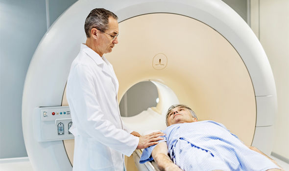
How Diagnostic Imaging Could One Day Affect Tax Laws for Personal Injury Settlements
With the 2017 passing of the Tax Cuts and Jobs Act, IRS tax policy on personal injury cases is back in the spotlight. At the same time, for the past few years, the legal profession has been absorbing the impact of ever-more detailed imaging studies that, some argue, display a physical basis for certain neurological phenomena, including disorders like PTSD and subjective experiences like pain itself.
Strangely, these two seemingly unrelated forces — tax laws for personal injury cases and the use of advanced imaging technologies in the courtroom — could combine to affect the take-home damages that plaintiffs receive from a successful lawsuit. Here's how, in a point-by-point analysis:
Tax laws for compensatory damages are complex, even circular. According to the IRS, damages paid out for physical injuries are tax-free. Damages for emotional injury, however, are fully taxable.
As researchers uncover more about the complex unity of physical and emotional systems in the human experience, courts and the IRS are faced with a challenge. Emotional trauma often leads to physical symptoms. Likewise, bodily injuries often result in emotional side effects. The two are difficult to separate. So which set of compensatory damages can legally go tax-free?
Under current precedent, the IRS holds that taxability is associated with the principal injury. Cascading effects are lumped in with the root complaint. So if a defendant physically injures or sickens the plaintiff, compensatory damages awarded in the case are tax-free, even if some of those damages are paid out for emotional effects associated with the injury. Conversely, if the defendant's negligence leads to emotional harm, and that psychological trauma manifests in physical symptoms, damages are fully taxable, even if some portion is awarded in compensation for bodily sickness arising from emotional distress.
The Tax Cuts and Jobs Act raises the stakes on this issue of taxation. Plaintiffs who hire contingent-fee attorneys are now responsible for paying taxes on 100 percent of case-related payments — including that portion that goes to pay the lawyer. Under previous law, attorneys and plaintiffs each assumed their share of the tax liability associated with taxable payments resulting from the same case.
Here's where advanced imaging could change the landscape. In 2008, the Arizona Superior Court in Pima County heard a landmark case, Koch v. Western Emulsions, Inc. The court allowed evidence of chronic neuropathic pain obtained with an fMRI scan, which the plaintiff's expert witness said showed a clear physical pattern of brain activity "proving" the existence of the pain. Ultimately, the defendant settled for $800,000, more than 10 times their initial offering.
Since Koch, the scientific community has warned against using advanced imaging as a sort of "painometer." The science just isn't there yet, and there are ethical issues involved in imposing "mind-reading" technologies on plaintiffs, researchers say.
But brain scans are showing physical changes associated with a lot more than pain. In one study, MRI scans showed changes in the physical structure of the hippocampus in Vietnam veterans with PTSD. Some had an 8 percent reduction in volume of the right hippocampus.
The question this all leads us to, then, is where to draw the line between physical and psychological injury. Advanced imaging technologies will continue to show greater levels of detail in the troubled brain. At some point, the courts and the IRS will have to wade into one of the most philosophically murky questions of all time: the mind-body problem. More to the point: Is there any injury, psychological or otherwise, that cannot be described as physical?
The answers we come up with will have dramatic effects on the value of settlements, damages, and tax payments in the years to come.
According to the American Medical Association (AMA) Code of Medical Ethics, "Managing health care resources responsibly for the benefit of all patients is compatible with physicians' primary obligation to serve the interests of individual patients." In other words, doctors should consider the cost of treatment, and save their patients money when they can.
At a time of record health care costs — more than $28,000 for the typical U.S. family in 2018 — this isn't just an issue of saving patients a few dollars here and there. When patients can't afford to pay their health care bills, they're more likely to delay seeking treatment. That delay can affect the outcome of eventual care.
Of course, as the AMA states, "Physicians' primary ethical obligation is to promote the well-being of individual patients." Sometimes that well-being hinges on cash or its lack. So how can general practitioners and other referring physicians limit the cost of care for their patients? Diagnostic imaging is a great place to start. The cost of imaging studies has grown faster than wages, overall inflation, and health care expense.
Luckily, doctors are pushing back against hospital pricing for these services. Here are a few ways referring physicians can provide excellent care for their patients without overspending on imaging studies:
Involve patients in the decision to seek or omit imaging tests. When a patient presents with conditions that aren't life threatening, doctors have significant leeway to work within the patient's preferences. Often, this leads to fewer imaging studies, with associated savings. In a recent study, doctors who used shared decision-making tools with their patients ordered 7 percent fewer advanced imaging tests and 30 percent fewer standard imaging studies.
Avoid ordering full-body scans to screen for tumors unless patients show symptoms. The American College of Preventive Medicine discourages the use of whole-body scanning to screen asymptomatic patients for tumors. They point out that no data suggests survival improvement for patients, and that less than 2 percent of asymptomatic patients screened had tumors.
Choose imaging providers with upfront pricing, and share that information with patients. It shouldn't be difficult to find pricing for imaging procedures before making a referral. If a provider conceals prices, choose another imaging clinic. With accurate pricing information in hand, physicians can work with patients to conduct a cost-benefit analysis of ordering a study.
Refer patients to high-quality imaging clinics rather than relying on hospital radiology departments. As we've mentioned in this space before, an MRI from a freestanding independent imaging center is often thousands of dollars less than the same procedure at a hospital — even with the same doctors and the same equipment. Choosing a high-quality imaging provider that's free from hospital pricing is the easiest way to save patients money on diagnostic tests.
Take advantage of digital delivery of diagnostic images and radiology reports. Doctors and patients can access digital reports anywhere and at any time, leading to lower costs and greater access. Precise Imaging offers doctors digital access and 24/7 tech support through a dedicated physician's portal.
With the ongoing public discussion of the U.S. health care system, awareness of the role of finances in treatment continues to rise. It's time to have these discussions; as of 2013, only 36 percent of surveyed physicians believed they had a "major responsibility" to control care costs on their patients' behalf.
But we know that affordability can translate into real-world effects on outcomes. Patient well-being and the costs of care are not two separate issues; they are bound together in complex, intractable ways. By choosing dedicated, patient-centered providers like Precise Imaging, doctors can get patients the services they need without unnecessarily adding to their burden of medical debt. Call us at 800-558-2223 or fill out our online form to make a referral today.
References:
Bradley D, Bradley K. The value of diagnostic medical imaging. North Carolina Medical Journal [serial online]. March 2014;75(2):121-125. Available from: MEDLINE Complete, Ipswich, MA. Accessed August 16, 2018.
Personal injury attorneys rely on MRI scans and other types of diagnostic imaging to build their cases. But not every hospital or clinic has the experience, the infrastructure, and the staff to provide crucial evidence when you need it.
Precise Imaging specializes in working with legal professionals while providing their clients with friendly, patient-centered service. We provide a single point-of-contact for management of all your personal injury cases, no matter how many you have at a time. We also have a designated web portal specifically for attorneys.
When you look for a diagnostic imaging center to work with, here are a few of the benefits you should look for — all of which you can find at Precise Imaging:
1. Patient-Centered Care
Diagnostic imaging built around the patient's health and comfort is at the core of Precise Imaging practice. Our radiologists, technologists, and support staff are all trained to communicate warmly and with respect for the patient's values and experience. We take cues, both verbal and nonverbal, and offer emotional support alongside physical comforts to ensure that patients can get back to their busy lives feeling good about the experience.
Patient satisfaction is always our goal. We understand that attorneys are motivated by their clients' success because we feel the same.
2. Flexible and Convenient Scheduling
Speaking of patient satisfaction, market research suggests that patients list scheduling difficulty and waiting time for appointments as major issues in their choice of health care provider. We suspect that attorneys who make appointments for their clients would say much the same.
With over 70 locations, Precise Imaging offers appointments for your clients when and where it's convenient for them. We can even schedule same-day imaging. Our radiologists provide reports within 24 hours of a scan — and faster when requested. Precise Imaging has years of experience working with the legal community, and we pride ourselves on taking the stress off patients and their representatives both through flexible scheduling.
When a client is struggling with an injury and a legal case, it helps to keep one thing, at least, as simple as possible. Our scheduling process is quick, easy, and convenient, no matter who makes the call.
3. HIPAA-Compliant Online Access to Images
Once a scan is completed and the radiologist analyzes the results, we offer images in the format of your choice. We can put the images on a CD or other digital device, or we can simply make them available to you, 24/7, through our attorney web portal.
The portal complies with all HIPAA regulations, allowing patients, health care professionals, and attorneys access to the information they need when they need it. Our attorney portal offers a user-friendly interface designed to simplify the organization of complex legal data.
4. Medical Lien Payments and Letters of Protection
Of course, unexpected medical bills can be difficult to cover. Your clients shouldn't worry about that while focusing on their cases. Precise Imaging accepts medical liens and letters of protection for personal injury to simplify patient finances during a trying time.
Precise Imaging offers a full suite of resources for attorneys. Find them here. And when you need MRI scans for personal injury lawsuits, contact Precise Imaging at 800-558-2223.
As evidence-based medicine (EBM) evolved into value-based medicine (VBM) — the "practice of medicine based upon the patient-perceived value conferred by an intervention," as Jong-Myon Bae defined it in the journal Epidemiology and Health — many referring physicians began to wonder about their choice of imaging providers.
Hospital-affiliated doctors can simply send a patient to the basement for an MRI or an ultrasound. But does this practice really contribute to a patient-centered model of care like VBM? Could physicians attain reliable diagnostic imaging at a reduced price for their patients, thereby balancing the outcome-to-cost ratio?
The answer is, resoundingly, yes. The reason? Independent imaging centers.
The price of an MRI at a freestanding clinic is often thousands of dollars less than the same scan, with the same outcomes, performed at a hospital. There are a few reasons for this — hospitals subsidize revenue-losing departments through radiology, and they're less vulnerable to market-driven competition — but suffice it to say, freestanding imaging centers provide the same service as hospitals at a fraction of the price.
In August of 2016, the European Society of Radiology's Working Group on Value-Based Imaging began its work investigating how radiology could better take part in the VBM model of patient care. Among the Working Group's findings:
The VBM model, as currently practiced, leaves diagnostic imaging out of the equation. Practitioners measure the success of value-based outcomes starting with treatment — which, notably, cannot begin until the radiologist has completed her role in an accurate diagnosis.
In fact, a traditional VBM approach only factors in diagnosis when it is incorrect, or when it causes complications. This bias neglects the value a patient places in a correct diagnosis, which is, the first in a string of health outcomes that truly matters to the patient and his family.
In order to fold radiology the VBM paradigm, imaging providers should measure and improve five process steps in their work: Determining whether the referral is appropriate, protecting patients from radiation, producing radiology reports that are entirely accurate and easy to understand, maintaining excellent relationships between patients and the provider's entire staff, and continuing education and innovation in the field.
Regarding that last point — arguably the Working Group's greatest revelation — note that freestanding imaging providers are often at the forefront of these five process steps. In a marketplace glutted with competition, no imaging provider can afford to fall behind in the quality of care they offer patients. And, as previously noted, freestanding imaging centers offer this care at a much lower price-point than traditional hospital-based radiology departments. Often, they employ the exact same radiologists as the hospitals, and operate identical equipment.
A true reckoning of value-based medicine starts with making the right diagnosis, and that often depends on great radiology. Independent imaging centers offer that level of quality at a lower price. Therefore, they are valuable partners in value-based care.
If you're a referring physician, the patient-centered approach is to talk to patients about their choice of diagnostic imaging providers. Often, the freestanding clinic leads to better outcome-to-cost ratios, which lie at the heart of value-based medicine.
Researchers at the Simmons Comprehensive Cancer Center have authored a study detailing a multiparametric magnetic resonance imaging (mpMRI) technique that predicts a malignant type of kidney cancer without performing a biopsy. The results of the method are impressive, but require more refinement to fully take the place of a biopsy.
Doctors frequently find kidney tumors accidentally while conducting CT scans for other reasons.
These scans alone do not yield the necessary information that tells doctors whether they are malignant or benign. Instead, a biopsy is usually performed. These procedures can save lives by correctly identifying the nature of the tumor, but they are also invasive and can cause complications.
“Using mpMRI, multiple types of images can be obtained from the renal mass and each one tells us something about the tissue,” Dr. Ivan Pedrosa, Professor of Radiology and Chief of Magnetic Resonance Imaging told theUT Southwestern Newsroom.
Identifying malignant masses in the kidney is extremely important because treatment is highly effective before the tumor metastasizes. However, once it spreads to other parts of the body, survival rates are low. Clear cell kidney carcinoma is an aggressive subtype of malignant masses that the researchers.
Seven radiologists studied the records of 110 patients with cT1a masses.
These patients had all undergone an MRI as well as a partial or radical nephrectomy. The observing radiologists did not know the final pathology findings, but instead relied on an algorithm to judge whether tumors were metastatic.
The researchers had 78 percent accuracy when rating that the mass was "probably" or "definitely" clear cell kidney carcinoma. When rating that the mass was possibly carcinoma, they had a 95 percent success rate.
The promising results show that biopsies may not be necessary for identifying certain cancers.
Because some patients are reluctant to consent to biopsies, this new technique is potentially lifesaving. As it stands, patients who do not want a biopsy may learn important information if an MRI shows that their kidney mass has a high probability of becoming metastatic. This new information could convince them that the pain of a biopsy is worth going through.
Using these methods to identify clear cell histology is still a work in progress. The doctors at Simmons Comprehensive Cancer Center will have to achieve a higher degree of accuracy in predicting malignant kidney masses for the method to become mainstream.
However, as standardization of imaging protocols and reporting criteria are refined, accurate results should increase. When MRI scans alone are sufficient to identify clear cell kidney carcinoma, doctors will have another powerful tool in the fight against cancer.
A first-time MRI procedure can make patients nervous, even to the point of ending the scan. That can lead to higher costs for imaging centers, and even affect patient outcomes if the anxiety interferes with the quality of the radiology report. Research shows one simple way to help nervous patients get through their scans without interruption: Communication.
A 2015 study in the journal Magnetic Resonance Imaging tested an intervention in which imaging staff explained the MRI process to one group of patients. They took blood samples during the scans, later testing them for the stress hormones prolactin and cortisol. Additionally, they took both the experimental and control groups through the 40-questions State-Trait Anxiety Inventory to measure patient nervousness.
The patients who had the intervention in which staff verbally shared information about the scan showed a 6-percent drop in cortisol after the scan. The control group's cortisol levels increased by 18 percent. The authors of the study conclude that "MRI anxiety can be reduced by information and communication. This combined method is shown to be effective and should be used during daily radiology routine."
So what can physicians do to prepare their patients for a first MRI scan in advance? It's never too early to start educating patients about what they can expect during a health procedure. And the MRI process can be boiled down into 10, easy-to-grasp steps. Share these steps with patients to help limit anxiety during an MRI scan:
First, radiology staff will walk the patient through a detailed screening process. Because of the strong magnetic field generated during an MRI, patients must report any medical implants or metal particles in their bodies. These may preclude the use of MRI imaging.
Once the patient clears the screening, staff will lead them into the MRI suite. Some imaging facilities offer hospital robes, to ensure there's no metal in the patient's clothing. Others allow patients to wear their own metal-free clothes, such as sweat pants and a T-shirt. The technologist will proceed to position the patient on the table; most commonly, patients lie on their backs. If the scan requires an additional radiofrequency coil, the technologist will place that on the patient's body at this time.
The patient enters the bore. Meanwhile, technologists cycle through a list of pre-programmed settings called "protocols." They'll choose the protocol that corresponds with the body part they are imaging; this will tell the MRI machine which angles, targets, and pulse sequences to use in this particular procedure.
Before the scan proper begins, technologists run a "scout" or "localizer" scan. This is a low-quality image, and it won't be used in reporting. However, localizer scans obtain visual and placement information that the computer will used to plan the angles of its imaging later in the process.
Parallel imaging is a process designed to speed up scan time. It collects less raw data during the scan, and patches missing information using special algorithms to generate the final image. Parallel imaging requires specifically calibrated coils, and may call for a calibration scan at this point.
One of the great strengths of MRI scans is that they create 3D images that can be viewed from any angle. The next step is to program in the angle of images for the radiologist. Technologists can change the "thickness" of the image at this point, as well.
Before the scanner can begin collecting valuable images, it must calibrate all systems through the use of a prescan. This shouldn't take much more than 10 or 20 seconds.
It is only at this relatively late stage in the process that the technologist actually runs the scan. They will make necessary adjustments and continue scanning according to the chosen protocol. In the end, they'll have clear, accurate images that radiologists will use in their reporting.
Some types of images require extra work in post-production, but this can be done after the patient has left the MRI suite.
Scanning complete, the technologist pulls the patient from the bore. Different types of scans take varying lengths of time, but most range between 20 and 60 minutes.
When patients understand more about their medical procedures, and know what to expect, they're less likely to experience significant anxiety. That's both a value in itself — as patient-centered caregivers, staff at Precise Imaging works to keep patients comfortable, both physically and emotionally — and an element of better diagnoses, which lead to better patient outcomes.
To learn more about an MRI procedure from Precise Imaging, or to refer a patient, call us at 800-558-2223.
References:
Deshmane A, Gulani V, Griswold MA, Seiberlich N. Parallel MR imaging. Journal of Magnetic Resonance Imaging: JMRI. 2012;36(1):55-72. doi:10.1002/jmri.23639
Tazegul G, Etcioglu E, Yildiz F, Tuney D. Can MRI related patient anxiety be prevented? Magnetic Resonance Imaging. 2015;33(1):180-3. doi:10.1016/j.mri.2014.08.024
Independent imaging centers are moving to the forefront of diagnostic care in the United States. Recently, at least one major health insurer dropped coverage for CT and MRI scans at hospital radiology facilities. The insurance company will only pay for these scans when patients visit providers who specialize in diagnostic imaging, and imaging alone.
How will this change the conversation between referring physicians and their patients? In order to explain the changes — why insurers would make a rule like this, and why it's not at all a bad change for patient care — physicians need only look at two key metrics of today's health care system: price and quality.
The Price Difference Between Hospitals and Independent Imaging Centers
When an insurance company makes a move like this, it's a clear indication that there's a wide price differential that's not necessarily associated with a difference in quality of care. Research from the Healthcare Financial Management Association — a professional organization for people who work on the financial side of the health care industry — shows that prices at hospitals are, in fact, dramatically higher than those at the average free-standing imaging provider. Among other differences, the HFMA found:
MRI scans cost an average of 70 percent more at hospitals than at independent imaging centers.
When those MRI scans covered the head and/or neck, they were an average of 80 percent more expensive in hospital-owned facilities.
The differences were even more stark for CT scans. Imaging of the body using CT cost 135 percent more at hospitals.
For CT scans of the head and/or neck, patients paid an average of 149 percent more at hospitals than at free-standing imaging facilities.
This research was published in 2017, though most of its data came from 2014. Either way, price gaps remains.
Measuring the Quality of Diagnostic Imaging Providers
Patients are used to associating higher prices with better service. In health care, however, quality and cost are independent of one another. The United States has the most expensive health care in the world.
In 2014, U.S. health care spending per capita was $9,237. That year the United Kingdom spent $3,749 per person while Japan's figure stood at $3,816. Still, among the 12 wealthiest industrialized nations — including Japan and the UK — the United States lands dead last in terms of life expectancy.
Clearly, spending more does not buy better care in this country. In fact, independent imaging centers offer measures of quality, in terms of better patient experience, that most hospitals cannot boast. At Precise Imaging, these include:
Evening and weekend hours to work around the patient's schedule, not the other way around.
Excellent, board-certified radiologists and technicians, often the same ones hospitals use, at a drastically reduced cost.
Quick reporting with HIPAA-compliant online sharing with referring physicians. Most doctors are reading radiology reports within 24 hours of the scan.
Same-day scheduling.
Transparent pricing.
There have been plenty of good reasons to refer patients to a free-standing imaging facility for years. Now that insurers are refusing to support the arbitrary and inflated costs that hospitals charge, more and more patients will be able to experience the care that Precise Imaging provides.
Patients and referring physicians increasingly enjoy the benefits of digital access to radiology reports, but the online environment raises new concerns about HIPAA compliance.
It is, of course, entirely appropriate for doctors to be concerned about patient privacy online. In May of 2017, at least 7,000 medical records were leaked at a New York City hospital. In November of that year, hackers took hostage the protected health information of 7,000 patients at a Massachusetts sports medicine provider, demanding a ransom with the threat of releasing the data. The following month, news broke that an ex-employee of a San Antonio mental health provider left the job with more than 28,000 patient records downloaded to a personal computer.
Data breaches happen, and when they do, health care providers could find themselves in violation of HIPAA. So how can physicians be sure that their radiology providers are adequately protecting their practices and their patients?
Here, we describe some of the ways that the Precise Imaging web portal does it. By following these recommended protocols, our IT systems keep out intruders, ensuring compliance with HIPAA to keep protected patient information safe and secure. First, though, we'll take a look at the letter of the law itself. What exactly does it mean for an online data-sharing system to be "HIPAA-compliant?"
The Health Insurance Portability and Accountability Act of 1996 and Patient Privacy
According to the HIPAA Security Rule, which governs the storage and transmission of electronic protected health information (EPHI), requires covered health care providers to meet four major goals:
The provider must keep EPHI in their possession — whether they create the information, hold it, or simply pass it along — completely private and confidential. It cannot become available to any non-approved parties.
Within reason it is the provider's responsibility to anticipate potential threats to secure information. If they identify a threat, they must act to protect EPHI from it.
Similarly, providers have a duty to shield EPHI within their control from "unauthorized uses or disclosures."
Finally, these requirements extend from top administrators to the very bottom of the pay scale. Every health care provider must make sure that all employees comply with the above rules.
Note that the Security Rule does not dictate specific technical steps companies must take to keep EPHI safe and secure. These things are up to the providers. And the entities covered under HIPAA have strong incentives for investing in ironclad digital protections for their patients' health information: They can face fines of up to $50,000 per compromised health record, with an annual maximum limit of $1.5 million.
But there's an even more important reason why responsible health care providers invest heavily in protecting patient information. Organizations that devote themselves to patient-centered care don't just treat a single injury or illness and forget their patients. They concern themselves with every aspect of the patient experience, from physical and emotional comfort to mental well-being between visits, as much that's possible. Finding out that your personal information has been leaked is a stressful experience, and no self-respecting health care provider wants to bring anxiety into a patient's life. In short, we invest heavily in privacy because that's what's best for the patient.
HIPAA Compliance in the Radiology Web Portal
Precise Imaging offers a series of user-specific web portals, for patients, referring physicians, and even personal injury attorneys who may need access to diagnostic images to win a case. Each of these web portals provides best-in-class security features, clearing HIPAA requirements and protecting our users' priceless data.
The following is far from an exhaustive list of the security tools that protect radiology reports and other EPHI within Precise Imaging web portals. But it should make clear that these systems are robustly protected from data loss that could put patient information at risk. Here are a few of the tools that our IT systems have in place to protect EPHI and comply fully with HIPAA requirements:
All transfers of EPHI through the Precise Imaging web portals are fully protected by SSL/TLS encryption.Transport Layer Security (TLS) is the industry standard for protecting data in transit from clients to servers and back again. This is an updated version of Secure Sockets Layer encryption (SSL), but we refer to the technology as "SSL/TLS" because people still tend to use the terms interchangeably. In fact, TLS is more advanced than SSL encryption, and that's what our web portals use to encrypt data in transit.
Data remains secure even in a computer's local cache through AES, the Advanced Encryption Standard. This block cipher algorithm is one of only two encryption tools used by the U.S. government, and it remains a powerful lock on data. To further protect data within the local cache, these web portals purge the cache of EPHI after each session. That is, after the user logs out, there's no patient information stored locally at all.
Site administrators set strict user identifiers. Each user must have a unique login ID and password, and our systems don't allow weak passwords. We may even ask for periodic password replacements; all of these systems are designed to keep EPHI as secure as possible.
Audit trail tools track each user's activities (without storing EPHI). If, somehow, an unauthorized user gained access to one of our web portals, integrated reporting tools would flag any suspicious activity.
After a period of inactivity, sessions will logout automatically. This reduces the chances of an unauthorized viewer gaining access to EPHI when users simply forget to log off.
The servers that Precise Imaging web portals use are configured specifically to comply with HIPAA. When it comes to patient data, we don't take chance. Server rooms that host patient data are even equipped with comprehensive physical security, greatly reducing the risk of intrusion.
There's no reason that compliance with HIPAA should reduce the web-based functionality that so many radiologists, referring physicians, and patients rely on to create better health outcomes. At Precise Imaging, we follow advanced security protocols to protect patient information. We're able to share online radiology reports with full HIPAA compliance, which tends to reassure even the most security-conscious referring physician we work with.
Gefen R, Bruno M, Abunedeh H. Online portals: Gateway to patient-centered radiology. American Journal of Roentgenology. 2017 209:5, 987-991. doi.org/10.2214/AJR.17.18291
Lee, CI, Langlotz CP, Elmore JG. Implications of Direct Patient Online Access to Radiology Reports Through Patient Web Portals. Journal of the American College of Radiology. 2016 13:12PB, 1608-1614. doi.org/10.1016/j.jacr.2016.09.007
Breast MRI is not a first-line breast cancer screening tool for most women. The American Cancer Society recommends annual MRI screening only for women with a lifetime risk of breast cancer that's greater than 20-25 percent, and only as an adjunct to mammography. The problem is that MR images are too clear, leading to too many false positives, the ACS explains.
"While the high rate of biopsies and further investigations is acceptable in women with a high risk of breast cancer, the number of such investigations in women at lower risk will be much higher than would be appropriate, leading to the need to counsel women in lower risk categories that MRI screening is not advisable and that the harms are believed to outweigh the benefits," wrote researchers for the American Cancer Society Breast Cancer Advisory Group in the ACS guidelines in 2007.
However, a study published in the May 2017 issue of the journal Radiology suggests that it could be time to rethink these recommendations. Researchers from the University of Aachen studied 2120 women, aged 40 to 70, with lifetime breast cancer risk factors of less than 15 percent. The women received screening via MRI, ultrasound, and mammography between 2005 and 2013.
Of the 60 cancers discovered in the study, 59 were observed using MRI scans. None were uncovered by ultrasound or mammography alone. Meanwhile, the positive predictive value of the MRI screening was high, hinting that concerns over false positives may be exaggerated given current MR technology and advances in radiology practice.
"In women at average risk for breast cancer, MR imaging screening improves early diagnosis of prognostically relevant breast cancer," the authors concluded.
Other Factors Limiting the Use of Breast MRI Screening
Even if further studies show that false positives are no longer as prevalent in MRI breast screening as they once were, other barriers continue to limit patient access to the procedure.
There are two major reasons physicians don't order more breast screening via MRI, reportsRadiology Today magazine: Insurance companies won't cover breast MRI for women unless they show high risk, citing excessive costs for the procedure; and there simply aren't enough MRI providers across the nation.
These are concerns that patients and physicians in many parts of California, Nevada, and Arizona needn't worry about. Precise Imaging operates a growing network of more than 70 free-standing imaging centers in communities across these states — with more on the way.
Precise Imaging Expands Access to Breast MRI Scans
By providing access to MRI scans within many communities, Precise Imaging allows patients to schedule appointments when and where they please. This greatly expands the total MRI capacity in areas where Precise Imaging operates.
As for the issue of cost, Precise Imaging provides a streamlined, flexible billing system, allowing patients to pay through as many avenues as possible. That includes Medicaid/Medicare, PPO insurance, personal injury liens, letters of protection, and low cash prices.
And because Precise Imaging providers aren't attached to a broader hospital system, they don't have to balloon prices to cover high-loss areas, such as emergency services. Together, these factors allow Precise Imaging to offer more diagnostic imaging procedures — including breast MRI — to more patients at a lower cost.
Some day soon, breast MRI might becomes a standard screening procedure for all women, with life-saving results.
References:
Kanal K, Butler P, Sengupta D, Bhargavan-Chatfield M, Coombs L, Morin R. U.S. Diagnostic Reference Levels and Achievable Doses for 10 Adult CT Examinations. Radiology. 2017 284:1, 120-133. doi:10.1148/radiol.2017161911
Saslow D, Boetes C, Burke W, et al. for the American Cancer Society Breast Cancer Advisory Group. American Cancer Society Guidelines for Breast Screening with MRI as an Adjunct to Mammography. CA: A Cancer Journal for Clinicians. 2007 57: 75–89. doi:10.3322/canjclin.57.2.75
Precise Imaging to Open New Anaheim Location, Expanding Availability of Patient-Centered Diagnostic Care with Unparalleled Access for Physicians
November 10, 2017 — Anaheim, California — Precise Imaging, a private corporation owned by Jaklin Benji, Matt Benji and Mike Rashidi, will open a new facility at 3174 West Lincoln Ave., in Southwest Anaheim, on the 13th day of November 2017. As a leading independent provider of diagnostic imaging services across California, Nevada, and Arizona this latest location will feature a state-of-the-art open MRI scanner. The opening of an Anaheim location will create even more flexibility for those in need of imaging services while expanding access for referring physicians and their patients.
"Access to quality care is our highest priority," said Danny Rackow, director of technology and operations at Precise Imaging. "The new Anaheim location gives doctors and patients yet another quality source for MRI procedures while bringing the benefits of an open MRI to a whole new community."
The Anaheim location adds to Precise Imaging's growing network of over 100 locations, many of which offer evening and weekend hours, same-day referrals, and 24-hour turnaround on radiology reports—even less for STAT cases.
Patient-Centered Care with Physician-Preferred Workflow
In keeping with Precise Imaging's mission to build dependable relationships with referring physicians in every medical specialty, the new location will offer a powerful suite of digital resources for health care providers. Physicians can make referrals at any time through a free online form. Following the procedure, they can view radiology reports and images 24 hours a day through a simple, HIPAA-compliant web portal. Experienced IT staff is available at every hour of the day and night to assist physicians with all digital tools.
"Doctors should be able to focus on care and not get bogged down on the simplest interactions such as scheduling, availability and ease of use with a provider," Mike Rashidi said. "We built our physician access tools to make the entire process hassle-free, from referral to reporting to interacting with insurance providers, because we want what the doctors want: better health outcomes for patients."
Precise Imaging has a proven track record of patient satisfaction, and the new location will continue this tradition. The inclusion of a comfortable open MRI scanner in the latest facility is just one example of the compassionate care that sets Precise Imaging apart. Friendly, experienced staff and board-certified radiologists take the time to provide support for the whole patient—body and mind—and that can lead to better, more actionable reports.
To learn more about Precise Imaging's new location, or to schedule an appointment anywhere in the network, call 800-558-2223.
About Precise Imaging
Precise Imaging is a leading provider of MRI, X-ray, PET, and other diagnostic services with locations across California, Arizona, and Nevada. The company offers industry-leading support for physicians and attorneys with best-in-class patient care. Flexible billing options include Medicaid/Medicare, PPO insurance, personal injury liens, cash prices, and more. To learn more about Precise Imaging, please visit www.precisemri.com.