According to the American Medical Association (AMA) Code of Medical Ethics, "Managing health care resources responsibly for the benefit of all patients is compatible with physicians' primary obligation to serve the interests of individual patients." In other words, doctors should consider the cost of treatment, and save their patients money when they can.
At a time of record health care costs — more than $28,000 for the typical U.S. family in 2018 — this isn't just an issue of saving patients a few dollars here and there. When patients can't afford to pay their health care bills, they're more likely to delay seeking treatment. That delay can affect the outcome of eventual care.
Of course, as the AMA states, "Physicians' primary ethical obligation is to promote the well-being of individual patients." Sometimes that well-being hinges on cash or its lack. So how can general practitioners and other referring physicians limit the cost of care for their patients? Diagnostic imaging is a great place to start. The cost of imaging studies has grown faster than wages, overall inflation, and health care expense.
Luckily, doctors are pushing back against hospital pricing for these services. Here are a few ways referring physicians can provide excellent care for their patients without overspending on imaging studies:
Involve patients in the decision to seek or omit imaging tests. When a patient presents with conditions that aren't life threatening, doctors have significant leeway to work within the patient's preferences. Often, this leads to fewer imaging studies, with associated savings. In a recent study, doctors who used shared decision-making tools with their patients ordered 7 percent fewer advanced imaging tests and 30 percent fewer standard imaging studies.
Avoid ordering full-body scans to screen for tumors unless patients show symptoms. The American College of Preventive Medicine discourages the use of whole-body scanning to screen asymptomatic patients for tumors. They point out that no data suggests survival improvement for patients, and that less than 2 percent of asymptomatic patients screened had tumors.
Choose imaging providers with upfront pricing, and share that information with patients. It shouldn't be difficult to find pricing for imaging procedures before making a referral. If a provider conceals prices, choose another imaging clinic. With accurate pricing information in hand, physicians can work with patients to conduct a cost-benefit analysis of ordering a study.
Refer patients to high-quality imaging clinics rather than relying on hospital radiology departments. As we've mentioned in this space before, an MRI from a freestanding independent imaging center is often thousands of dollars less than the same procedure at a hospital — even with the same doctors and the same equipment. Choosing a high-quality imaging provider that's free from hospital pricing is the easiest way to save patients money on diagnostic tests.
Take advantage of digital delivery of diagnostic images and radiology reports. Doctors and patients can access digital reports anywhere and at any time, leading to lower costs and greater access. Precise Imaging offers doctors digital access and 24/7 tech support through a dedicated physician's portal.
With the ongoing public discussion of the U.S. health care system, awareness of the role of finances in treatment continues to rise. It's time to have these discussions; as of 2013, only 36 percent of surveyed physicians believed they had a "major responsibility" to control care costs on their patients' behalf.
But we know that affordability can translate into real-world effects on outcomes. Patient well-being and the costs of care are not two separate issues; they are bound together in complex, intractable ways. By choosing dedicated, patient-centered providers like Precise Imaging, doctors can get patients the services they need without unnecessarily adding to their burden of medical debt. Call us at 800-558-2223 or fill out our online form to make a referral today.
References:
Bradley D, Bradley K. The value of diagnostic medical imaging. North Carolina Medical Journal [serial online]. March 2014;75(2):121-125. Available from: MEDLINE Complete, Ipswich, MA. Accessed August 16, 2018.
Traumatic brain injury (TBI) can be one of the most devastating results of an at-fault accident, in a car, on the job, or even just walking down the sidewalk. The sheer volume of human systems that the brain controls — all of them, essentially — leads to an extraordinarily diverse set of symptoms in cases of TBI. Convincing judges and jurors that these disparate symptoms can all be traced back to a preventable injury is not always easy.
The irony here is that a moderate or severe TBI can lead to challenges that most deserve restitution. Some brain injuries are so severe that they deprive the victim of the ability to work. Others might even prevent a patient from participating in the tasks of daily living necessary for independence. If a client in a TBI-related personal-injury case is to be made whole, settlements may have to provide for them for the rest of their lives.
This combination of high stakes and soft proofs makes it imperative that attorneys in such cases understand diagnostic imaging. After all, while a defense team may be able to convince the judge that behavior changes are unrelated to an accident, it is hard to argue with an image of a damaged brain.
Here are a few things that personal injury lawyers should know about diagnostic imaging as it relates to TBI:
Different modalities excel at documenting different types of injuries. Radiologists are likely to use standard diagnostic tests like CT and MRI scans to diagnose TBI. They might order X-ray scans, but only in cases of suspected damage to the skull; X-ray images do not differentiate between soft tissues. Doctors might even prescribe advanced imaging techniques, such as perfusion CT, diffusion tensor imaging (DTI), or magnetoencephalography, a form of fMRI.
CT scans without contrast are the primary modality used to identify most primary, acute injuries. A TBI is not a single injury, but a cluster of related events. Doctors divide TBI into primary and secondary injuries. The first occurs at the moment of impact, causing a chain-reaction of events within the brain that often lead to further damage: secondary injuries. This is why doctors typically prescribe ongoing diagnostic imaging for TBI patients.
While advanced imaging modalities including brain-function tests like PET scans and functional MRI may identify secondary injuries, the first-line test for diagnosing primary TBI is the noncontrast CT scan. This is the fastest way to accurately expose bleeding that requires immediate surgery.
Doctors use MRI scans to identify certain types of TBIs that do not always show on a CT scan. While doctors typically order CT scans first, there are some types of TBI that require MRI for identification. When a primary TBI does not result in intracranial bleeding, MRI scans are often the better choice. Injuries that the MRI identifies better than CT scans include brain bruising and traumatic axonal injury (TAI), shear-strain damage to white matter caused by the brain's rapid acceleration within the skull.
As in any case involving diagnostic images, attorneys should engage expert witnesses such as neurologists or radiologists to explain what's going on in the pictures. This is particularly important in brain-function scans, such as fMRI and PET scans, which are often inscrutable for untrained viewers.
The good news for attorneys new to TBI cases and the diagnostic interventions that document them is that expert help is available. This brings us to our next point.
Expert Assistance for Personal Injury Cases Representing Victims of TBI
Personal injury lawyers and their clients deserve the chance to focus entirely on a TBI case. That can't happen when they're navigating complex scheduling systems at large hospitals or arguing with insurance companies. That's why Precise MRI always strives to provide for attorneys and their clients with what they need, when they need it.
Precise MRI offers quick turnarounds on radiology reports, with same-day scans and results within 24 hours. We provide more than 70 imaging centers in California, Arizona, and Nevada for convenient, close-to-home access for patients. Our friendly scheduling staff will find an appointment that works for your client, even on weekends and evenings.
Attorneys themselves have their client's crucial information at their fingertips thanks to an online portal designed for legal professionals. It's available 24/7, and so is IT service, ensuring that patients and their lawyers can access medical data at their own convenience.
We accept personal injury liens — and even offer a free, downloadable lien form for immediate access — and attorney letters of protection for personal injury. Our teams of fully certified medical professionals have long-term experience working with attorneys on all sorts of personal injury cases, including those involving TBI. Even more important, they're devoted to a patient-based model of care, and work hard to ensure quality, comfort, and convenience for all.
To learn more about attorney resources from Precise Imaging, or to schedule a CT or MRI scan for a client, call us today at 800-558-2223.
References:
Hill CS, Coleman MP, Menon DK. Traumatic Axonal Injury: Mechanisms and Translational Opportunities. Trends in Neurosciences. 2016;39(5):311-324. doi:10.1016/j.tins.2016.03.002.
Kim JJ, Gean AD. Imaging for the Diagnosis and Management of Traumatic Brain Injury. Neurotherapeutics. 2011;8(1):39-53. doi:10.1007/s13311-010-0003-3.
There are currently 5.7 million people living with Alzheimer's disease (AD) in the United States, and this number is projected to reach 14 million by the year 2020. As of 2018, AD was the fifth-leading cause of death of seniors, taking more lives than breast or prostate cancer combined. With such staggering and ever-increasing numbers, the need for early detection and treatment has reached a crisis point.
But understanding AD hasn’t been easy. Researchers have struggled to identify the true cause of this disease, develop a standard treatment plan, or find a cure. While an AD diagnosis may seem dire, early detection of the disease can help identify how the disease progresses, which in turn can help to create a treatment option.
New Research on AD Biomarkers and How to Detect Them
Recent research has shown a connection between changes in the brain’s anatomy and biomarkers known to appear at the early signs of AD. These biomarkers occur before any sign of cognitive problems, meaning these markers could possibly lead to a new, non-invasive AD screening test. Researchers have already discovered that the build up of amyloid-Beta and tau proteins on the brain, as well as a loss of volume in the hippocampus, are early signs of AD.
To further examine any links between these two phenomena, researchers from McGill University and McGill-affiliated health institutes studied 88 AD at-risk individuals with no signs of any cognitive decline from the disease. The subjects were given MRI scans to check brain volume, and also had cerebrospinal fluid samples taken to test levels of amyloid-Beta and tau proteins.
The researchers found that high levels of both amyloid-Beta and tau proteins were associated with loss of hippocampus volume, but there was no loss in volume when only one of the proteins accumulates within the brain. This suggests that doctors may someday be able to use MRI scans to monitor changes in the brains of AD patients at a microstructural level, before more serious changes begin to take place.
The recognition that symptoms of AD progress from physiological to cognitive can help diagnose those most at risk of developing this disease. These biomarkers might also help with testing the effectiveness of trial medications, and might one day allow physicians to target at-risk individuals with a simply MRI scan rather than a painful lumbar puncture.
The Expanding Role of MRI Scans in AD Diagnosis and Treatment
Non-invasive tests for AD have ramifications that extend well beyond patients themselves, to friends, family, and society at large. The fact is, in addition to being heartbreaking, Alzheimer’s is an incredibly costly disease. Many AD patients require more hospital visits, as well as full-time, long term care. However, accurate and early diagnosis of Alzheimer’s disease could help save $7.9 trillion is medical costs—and relieve some strain on family members and friends.
Thanks to MRI scans and their potential role in spotting Alzheimer's before cognitive symptoms appear, we seem to be edging ever-closer to the ultimate goal of treating AD effectively.
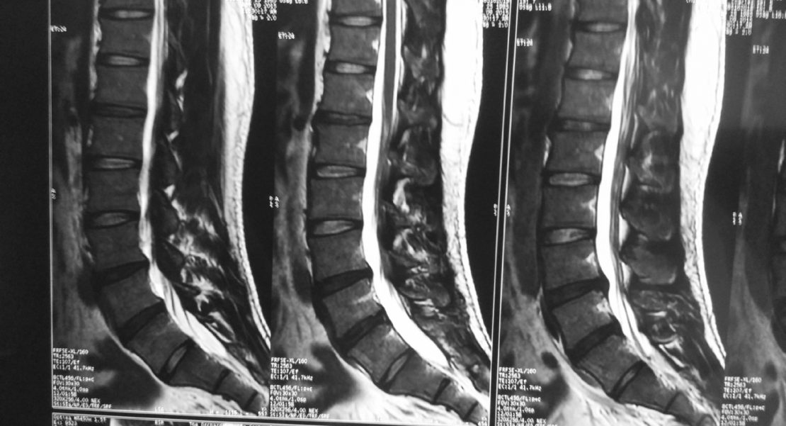
It's difficult to overstate the importance of diagnostic images in spinal injury litigation. When properly presented, a diagnostic image offers clear, compelling evidence—but if the image doesn't clearly show the injury, or if there's any question as to the validity of the attorney's interpretation, the entire case can be compromised. Of course, every personal injury attorney understands these points, but in order to use images effectively, they should understand the basics of different imaging technologies. That includes the tactics that radiology professionals must employ to deliver high-quality, diagnostically relevant images.
How Types of Spinal Injuries Affect the Diagnostic Imaging Process
The first consideration is the severity and location of the injury. Magnetic resonance imaging (MRI) provides the best diagnostic images for spinal cord and soft-tissue injuries, both for radiology professionals and for laypersons (while diagnostic images need to be evaluated by a trained professional, the images are obviously more useful to attorneys if the injuries are clearly visible and easy to explain). MRIs can show chronic issues such as spinal misalignment. Spinal fractures are easier to identify via computed tomography (CT), while spinal vascular injuries can be evaluated with either technology. In any case, trained radiology professionals working with the latest tech can discern a surprising amount of information, including the approximate age of the injury, which can help attorneys build winning cases.
Choosing Diagnostic Imaging Services for Personal Injury Cases Personal injury attorneys should keep other practical considerations in mind when directing their clients. As mentioned earlier, timing is often a crucial factor, particularly in spinal fracture cases. An imaging facility should be able to produce high-quality results quickly and at a fair cost — and, for the benefit of the attorney, those images should be in commonly used digital formats, ready for reproduction. Many attorneys choose to highlight certain parts of MRIs and CT scans with software designed for the purpose; without exceptional images, this can be a difficult process.
Finally, as we've mentioned on other blogs, imaging centers should be prepared to handle various types of remuneration and should have experience with workers' compensation and letters of protection.
Precise Imaging offers dedicated tools for attorneys through a specialized HIPAA-compliant web portal, which provides 24/7 access to images, case details, and payment information. We're dedicated to keeping patients comfortable, and with more than 70 facilities, we offer unequaled access to qualified imaging experts and state-of-the-art technology. To learn more about Precise Imaging’s commitment to assisting in personal injury cases, or to refer a client today, call 800.558.2223. You can also make an online referral here.
References:
Goldberg AL, Kershah SM. Advances in Imaging of Vertebral and Spinal Cord Injury. The Journal of Spinal Cord Medicine. 2010;33(2):105-116. PMID: 20486529
Parizel PM, van der Zijden T, Gaudino S, et al. Trauma of the spine and spinal cord: imaging strategies. European Spine Journal. 2010;19(Suppl 1):8-17. doi:10.1007/s00586-009-1123-5.
Ellingson BM, Salamon N, Holly LT. Imaging Techniques in Spinal Cord Injury. World neurosurgery. 2014;82(6):1351-1358. doi:10.1016/j.wneu.2012.12.004.
Diagnostic Imaging in The AMA's Guides to the Evaluation of Permanent Impairment
If you had to pick one book to be the authority on assessing personal damage in U.S. tort-civil law, you might choose The American Medical Association's Guides to the Evaluation of Permanent Impairment (AMA Guides). Personal injury law is more complicated than a single book could encompass, of course, but to this day, many U.S. courts rely on the systems set forth in the AMA Guides to determine a "quantitative estimate of function losses" within a victim's life. Many states use the AMA Guides to settle questions of workers' compensation and disability claims.
The AMA Guides assessment can also be a powerful tool in a personal injury case.
Note that the types of losses evaluated in the Guides may lead to pecuniary and/or non-pecuniary damages. In the former category, the Guides systems might uncover a loss of earning capacity or, in cases of disability, the necessity of long-term care. The AMA's Guides to the Evaluation of Permanent Impairment may also play into a claim of quality-of-life loss or limited ability due to the injury, both non-pecuniary damages.
As we've mentioned in previous posts, diagnostic imaging can also occupy a major role in personal injury cases. So the question is: How do The AMA's Guides to the Evaluation of Permanent Impairment implement diagnostic imaging as a factor in their determination systems? And which modalities most clearly demonstrate injury, according to the Guides?
Here are a few key facts about the intersection of diagnostic imaging and The AMA's Guides to the Evaluation of Permanent Impairment that every personal injury attorney should know:
Diagnostic Imaging Studies are an Important Part of Injury Assessment in the AMA Guides to the Evaluation of Permanent Impairment
In assessing impairment or disability, as in health care itself, diagnostic imaging plays an outsized role. The AMA Guides present each body system in its own chapter, introducing principles of assessment for each system. In nearly every chapter, some form of diagnostic imaging appears in the Guides' assessment criteria.
The results of all diagnostic studies — including imaging — must also be part of the physician's report, according to the Guides.
Imaging Modalities May Differ from One Edition to the Next
Editions matter. Depending which state you're in, the worker's compensation code may defer to the 6th and latest edition of the AMA Guides — or it might not. Some states use the 5th or even earlier editions, while others prefer state-specific criteria for evaluating injury, unrelated to the Guides. And while these codes relate specifically to worker's compensation rather than personal injury cases, the court's familiarity with a particular edition could add up to more compelling evidence of preventable injury and lasting impairment.
Differences between editions complicate the evaluating physician's choice of imaging modality. For instance, according to analysis from health care news site Medscape, methodology changes between the 5th and 6th editions of the Guides upend the rules for assessing impairment in a knee joint injury.
"No provision was made for ratings based on magnetic resonance imaging (MRI) or arthroscopic findings of cartilage pathology in the 5th edition of the Guides," reports Medscape. "However, in the 6th edition of the Guides, there is provision for full-thickness articular cartilage defects, with a range of 5 to 9 percent impairment...MRI and arthroscopy are objective measures with accepted grading systems for cartilage lesions."
So should an attorney ask the evaluating physician for an X-ray or an MRI scan in a knee-injury case? It may depend on which court tries the case.
Imaging Findings Alone are Not Enough
The 5th Edition of the AMA Guides makes the important point that the results of an imaging study alone aren't enough to classify an injury as impairment or disability.
The purpose of an assessment using AMA Guides systems is to establish a diagnosis-related estimate, or DRE. DRE categories are a measure of the "impairment of the whole person," expressed as a percentage range, with 0 percent impairment equating to no injury and 100 percent representing imminent mortality.
"To be of diagnostic value, clinical symptoms and signs must agree with the imaging findings," the 5th edition AMA Guides claim. "In other words, an imaging test is useful to confirm a diagnosis, but an imaging result alone is insufficient to qualify for a DRE category."
So where does all of this leave personal injury attorneys considering the inclusion of diagnostic imaging into a particular case? The key is to work as closely as possible with the client's attending physician or, in some cases, a court-approved health care provider. In the medical setting, imaging is a crucial part of diagnosis, which is necessary for healing. In a court of law, radiographs and MRI scans are just as valuable — but for the very different purpose of assessing injury for a fair damage claim.
Interested attorneys can purchase a copy of The American Medical Association's Guides to the Evaluation of Permanent Impairment at the AMA webstore, here. For more information on how Precise Imaging can help personal injury firms better serve their clients, see our Attorney Resources page, or contact us at 800-558-2223 today.
Personal injury liens help victims obtain medical treatment and stay afloat financially while their case is being decided. While this agreement entitles medical centers to repayment after a case has been settled, it can be greatly beneficial to attorneys and their clients as well.
Because many people cannot afford to pay major unforeseen medical bills, liens provide a lifeline until a settlement is reached, and attorneys benefit by ensuring that their clients get medical procedures and imaging completed in a timely manner. Still, there are a few things to consider when advising clients on this complicated subject.
The first thing to consider is what kind of experience the health care facility has with liens. Many smaller medical centers and imaging facilities don't accept personal injury liens; others may have a poor history of working out reimbursements. While the client may not have a choice of their initial point of care, they can certainly choose where subsequent imaging is done.
And that medical imaging is important for not only the person's future medical care, but also the outcome of their personal injury case.
Magnetic resonance imaging (MRI) scans can make or break a case.
Some physicians are reluctant to order MRI scans for accident victims — they feel that the scanning technology is so good at detecting problems that it can reveal false positives through asymptomatic abnormalities. However, there are many reasons that American courts encourage MRI scans.
For one, an MRI scan is completely objective. While radiologists may have differing interpretations of the scans, the scans themselves cannot be manipulated to distort the truth. That makes for powerful evidence to a judge or jury. Seeing the results of negligence in a black and white scan trumps many other forms of evidence.
Furthermore, seeing clear evidence of an injury can increase the value of a claim. Injuries in the brain, spinal discs, nerves, and joints can be difficult to verify without this state-of-the-art imagery. An MRI shows what many other forms of imaging cannot, and that's makes it invaluable to winning and maximizing personal injury lawsuits.
Of course, some patients may be reluctant to get MRI scans for a variety of reasons.
In these cases, victims of injury should be aware of how beneficial these scans can be for correct diagnosis and beginning recovery (not to mention, winning a case). Magnetic resonance allows doctors to pinpoint the source of an injury with the very latest technology available.
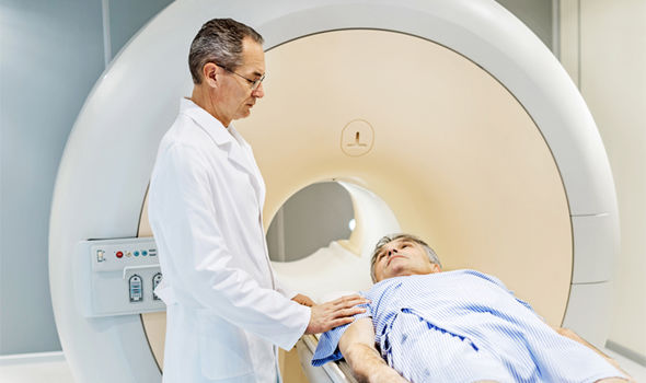
Some people hesitate to complete scans because of anxiety over claustrophobia. These fears are completely understandable, but there has been progress in recent years in making the technology more patient-friendly. For instance, open MRIs can help keep patients calm by eliminating the closed spaces that exist in traditional MRI machines. Even simple strategies, like prescribing an anti-anxiety medication or providing headphones with music can help anxious patients get through a scan.
Choosing an imaging center with personal injury experience can make the process easier for patients and attorneys.
The biggest reason is that facilities with experience offer a patient-centered focus and streamlined approach to accessing medical images. These facilities understand HIPAA regulations, know the preferred formats for images, and offer web portals for easy accessibility.
Using a single, preferred diagnostic company can also simplify caseloads for attorneys.
By relying on a trusted imaging center, firms know that crucial evidence is always just a click away — no matter what time of day. Attorneys can also trust that their clients are getting the best rates, so that injury victims can hold onto the lion's share of their settlement.
Finding a diagnostic imaging center that can handle time-sensitive requests is also important. Many hospitals experience lengthy delays in scheduling scans. Because Precise Imaging has over 70 locations, we can schedule same-day scans. Even better, our radiologists will file reports no later than 48 hours after the scan (and sometimes much sooner). The quick scheduling and prompt reading allows personal injury cases to clear significant hurdles without taking up too much time.
Look for an imaging center with web portals for attorneys.
Because legal cases are time-sensitive, having a 24-7 web portal available is crucial. Precise Imaging provides such portals so that attorneys can access important information night or day. Our web portals provide medical images, payment information, and case details, all while fully complying with HIPAA regulations. This service keeps attorneys organized and on track without having to rely on the medical center being open for service.
Our facilities are well-acquainted with the needs of personal injury lawyers, and our patient-centered facilities can comfort distraught patients who have recently suffered injuries. We offer a number of anxiety-reducing strategies to make our MRI scans as comfortable as possible.
We can schedule scans for the same day and get you results within 48 hours. All of the information an attorney or patient needs to access can be found on our 24-hour-per-day web portal. These services simplify a personal injury case and keep you on track.
We have partnered with patients and attorneys for thousands of personal injury cases and take our responsibilities to all parties seriously. We are prepared to accept all forms of payment, including workers' comp, liens, and deferred payment (through a letter of protection).
Precise Imaging makes scans fast and easy for patients and provides streamlined results for attorneys. If you'd like to partner with Precise Imaging for your personal injury cases, call 800.558.2223 or make an online referral here.
Magnetic resonance imaging (MRI) scans can be crucial pieces of evidence in personal injury lawsuits. The detailed images can convince a jury or judge that an injury occurred as a result of an accident or negligence, rather than aging and genetics.
MRI scans are universally accepted by insurance companies and courts because of their objectivity and superiority to older forms of imaging, like X-ray. Different radiologists may have differing interpretations of the scans, but in general, the results of an MRI scan provide definitive evidence to a judge or jury.
Another great advantage of MRI scans is that they use no radiation, and thus are safer than alternative forms of medical imaging. Instead, MRI scans use powerful magnets to develop highly detailed scans of the body. These powerful magnets require special attention for those with pacemakers or other implanted devices, but scans can still be safely given to most cardiac patients.
Litigation MRI scans are helping everyone from injured motorists to football players with traumatic brain injuries. It's this versatility that makes MRI scans the gold standard for determining medical history.
Unfortunately, MRI scanners at hospitals are expensive and in high-demand.
This may lead the doctor to prescribe rest for your client's injury before signing off on an MRI scan. If you'd like to move your case along, we can help schedule an affordable and timely scan. Precise Imaging has years of experience and accepts cases on lien. Advantages of working with us include:
Fast and easy scheduling - Call Precise Imaging at (800) 558-2223 or schedule an appointment online. We have over 70 locations, many open nights and weekends, to get your scans done quickly and professionally.
A variety of delivery options - Obtain scans on compact discs or other secure media for reliable access. We're experienced in handling potential evidence and same-day reporting is available in many locations.
A range of imaging modalities - Whether you need dual-focus X-ray tubes or an open MRI machine, we've got you covered. For any type of medical imaging, we'll harness our flexibility to get you the best images for your case.
Our facilities are patient-focused - We know that patients who have suffered an injury are under stress and possibly in pain. Our highly trained techs will make sure patients are comfortable and stress-free as they undergo imaging.
Precise Imaging has served more than 150,000 patients throughout California, Nevada, and Arizona. Whether you're an attorney or a patient, we can schedule an appointment at a convenient time and place. Call (800) 558-2223 or make an appointment online.
Preparing Patients to Read Their Own Radiology Reports
Increasingly, diagnostic imaging providers use web portals to give patients full access to their own radiology reports. Despite early fears that patients would misinterpret complex medical information, leading to unnecessary anxiety, most studies of direct access to radiology reports suggest that this is a positive step for patients and health care providers alike. When patients take a more active role in their care, they provide an extra level of oversight, reducing the chance of an error. Informed patients are also more likely to ask radiologists helpful questions, potentially preventing unnecessary testing.
In order to realize the full benefits of open access, however, primary care physicians or radiologists themselves should start educating their patients on how to read radiology reports. Otherwise, the fears of the opponents of patient access are more likely to be borne out in the patient population.
Here are a few of the key points to emphasize to patients who may be accessing their radiology reports for the first time:
Explaining Radiology Reports to Patients: The Breakdown
Patients may be more comfortable with the radiology report when you explain it section-by-section. Luckily, radiology reports are broken down into six sections, which is a good place to start when explaining the documents to patients. Let patients know that the typical radiology report will include the following: The type of exam performed, the patient's clinical history, the comparison, the imaging technique, findings, and the radiologist's final impressions. Encourage patients to ask questions if they encounter anything they don't understand in any of these six sections:
Section 1: The Basics
Radiology reports that begin with naming the type of exam will explain which imaging modality (MRI, CT, X-ray, etc.) was used in the procedure — and, crucially, which part of the body technologists scanned. They'll also include the time and date, as well as any details about preliminary procedures, like the use of contrast.
When discussing this first section, make sure patients understand the basics of the modality, and risks involved. This is a good time, for instance, to mention that MRI scans don't expose patients to ionizing radiation, or that intravenous contrast has a low incidence of negative side effects.
Section 2: The Patient's Backstory
The “clinical history” section of a report includes information such as the patient’s age, sex, and pre-existing conditions or diseases, and any other relevant medical information needed.
Patients can find the reason for the scan and, often, a suspected diagnosis in this section. This information helps the radiologist focus the report to each patient’s individual medical situation. Conversations with patients about their clinical histories can ensure that physicians have access to all relevant data before referring the patient for diagnostic imaging.
Section 3: Comparing New Scans to Previous Radiology Reports
If the patient has had other imaging done in the past, sometimes those images need to be used as comparisons. The “comparison” section of the typical radiology report is where past images will be mentioned. Typically, radiologists only consult scans of similar regions of the body when making comparison notes.
Section 4: Technical Details
When patients have questions about a radiology report, they often turn up in the "technique" section. Ironically, the details provided here — exact scientific descriptions of the imaging procedure — may not give the referring physician or the patient strongly relevant information.
However, the information contained herein is vital for radiologists, who may need to recreate or alter technical procedures in later scans. In reading through the technique portion of the radiology report, physicians may need to explain medical terminology, such as the use of anatomical planes, as well as structures of the body.
Section 5: The Radiologist's Findings
This is what every patient wants to know—what was found during a scan. Don't let patients place too much emphasis on the findings, however; the detailed analysis comes with the radiologist's impressions, in the final section of the typical radiology report.
Here, radiologists provide their notes on the normality or abnormality of the scanned area. Sometimes, patients don't see a certain organ or bodily structure that they know was part of the imaging procedure. Let them know that this usually just means the radiologist didn't find anything worth commenting on in that area. That's usually good news.
Section 6: The Radiologist's Diagnosis
The "impression" section of a medical report is where patients will find the radiologist's diagnosis, along with recommendations for future testing for confirmation. Commonly, radiologists offer differential diagnoses, which include any number of potential causes of the patient's symptoms. This can be confusing to patients new to reading radiology reports, so it can be helpful to spend extra time discussing each of the diagnoses on the radiologist's list, and planning for future diagnostics that can uncover the true illness.
The Growth of Portal-Based Radiology Reporting
Even if the diagnostic imaging facilities you refer patients to don't offer online access to radiology reports, odds are they will in the near future. The latest survey found that nearly 80 percent of patients who responded said they preferred to view radiology reports through online portals compared with more traditional methods of reporting. Tellingly, this includes getting the details verbally from their referring physician.
Another survey of 617 patients found that 64 percent of the study participants said they wanted to review their radiology reports themselves. An impressive 85 percent reported a desire to see the diagnostic images themselves.
In a consumer-driven health care market, these preferences are bound to win out. Prepare patients to read their own radiology reports to improve the efficacy of patient web portals at diagnostic imaging centers.
References:
Cabarrus, M et al. Patients Prefer Results From the Ordering Provider and Access to Their Radiology Reports. Journal of the American College of Radiology. June 2015; 12(6):556-562. DOI: https://doi.org/10.1016/j.jacr.2014.12.009. Accessed March 14, 2018.
Johnson, AJ et al. Access to Radiologic Reports via a Patient Portal: Clinical Simulations to Investigate Patient Preferences. Journal of the American College of Radiology. April 2012; 9(4):256-263. DOI: https://doi.org/10.1016/j.jacr.2011.12.023. Accessed March 14, 2018.
Landro L. Radiologists Push for Medical Reports Patients Can Understand. The Wall Street Journal. September 2014; Web. Available here. Accessed March 14, 2018.
Orenstein B. Reporting to Patients. Radiology Today. January 2013;14(1):22. Available here. Accessed March 14, 2018.
Patients accessing medical records online more than ever. Becker's Hospital Review. July 2016; Web. Available here. Accessed March 14, 2018.
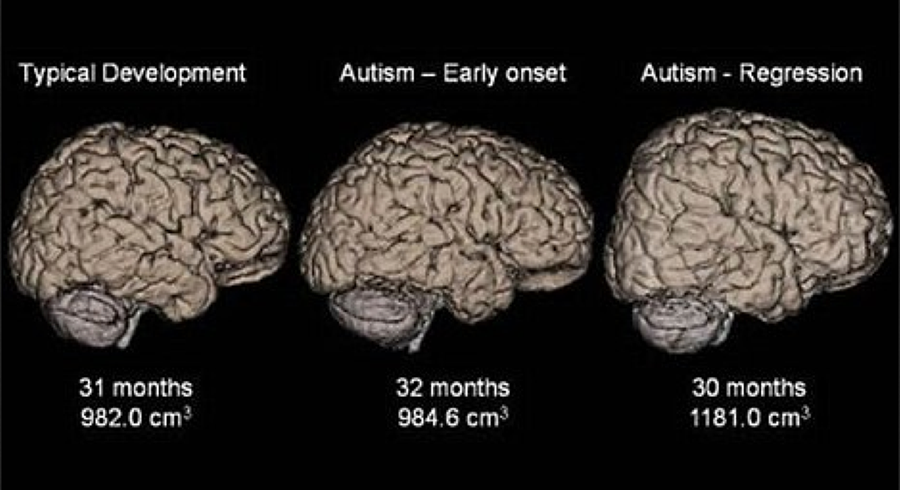
Researchers have observed abnormal neural networks in preschoolers with autism spectrum disorder (ASD) using a specialized magnetic resonance imaging (MRI) technique. The discovery gives hope that MRI scanning may one day allow early diagnosis, intervention, and treatment in ASD.
The study included 21 children with ASD and 21 children with typical development (TD).
It took place over four years in the Chinese PLA Hospital in Beijing. The researchers used a special MRI technique called diffusion-tensor imaging (DTI), which allowed them to observe the location, orientation, and anisotropy of white matter tracts in the children's brains.
Other scientists have used a similar technique to study the brains of patients with Alzheimer disease, multiple sclerosis, epilepsy, and other psychiatric disorders. The approach has led to
an improved understanding of topological organization of the brain network in patients with these disorders.
There were similarly useful results in the study of preschoolers with ASD. The researchers observed alterations of white matter in the children with ASD compared to children with TD. The scientists were able to correlate the alterations in the white matter networks with delays in verbal communication, object use, visual response, body use, and listening response.
The study also observed increased nodal efficiency in children with ASD compared with TD.
That observation agreed with previous studies that had observed this phenomenon in adults on the autism spectrum. The researchers believe this is a reflection of a delayed maturation process in people with ASD.
"Altered brain connectivity may be a key pathophysiological feature of ASD," study co-author Lin Ma told Science Daily. "This altered connectivity is visualized in our findings, thus providing a further step in understanding ASD. The imaging finding of those 'targets' may be a clue for future diagnosis and even for therapeutic intervention in preschool children with ASD."
Medical professionals from around the world weighed in on the study.
"This discovery gives us a more objective diagnostic method by using MRIs to aid us in the diagnosis of children who do have autism, and also gives us a better understanding of the abnormal differences in the brain," psychiatrist Dr. Matthew Lorber told HealthDay Reporter.
Doctors presently diagnose ASD through observing difficulties with social use of communication and interaction in children. A standardized MRI technique that could identify the disorder could give parents and therapists an earlier chance at intervention and treatment.
It's still too early to rely on MRI scans for the diagnosis of autism, but the study's results show that it's possible. The study also provides other researchers with imaging biomarkers that may speed the development of this technology.
Researchers at the Simmons Comprehensive Cancer Center have authored a study detailing a multiparametric magnetic resonance imaging (mpMRI) technique that predicts a malignant type of kidney cancer without performing a biopsy. The results of the method are impressive, but require more refinement to fully take the place of a biopsy.
Doctors frequently find kidney tumors accidentally while conducting CT scans for other reasons.
These scans alone do not yield the necessary information that tells doctors whether they are malignant or benign. Instead, a biopsy is usually performed. These procedures can save lives by correctly identifying the nature of the tumor, but they are also invasive and can cause complications.
“Using mpMRI, multiple types of images can be obtained from the renal mass and each one tells us something about the tissue,” Dr. Ivan Pedrosa, Professor of Radiology and Chief of Magnetic Resonance Imaging told theUT Southwestern Newsroom.
Identifying malignant masses in the kidney is extremely important because treatment is highly effective before the tumor metastasizes. However, once it spreads to other parts of the body, survival rates are low. Clear cell kidney carcinoma is an aggressive subtype of malignant masses that the researchers.
Seven radiologists studied the records of 110 patients with cT1a masses.
These patients had all undergone an MRI as well as a partial or radical nephrectomy. The observing radiologists did not know the final pathology findings, but instead relied on an algorithm to judge whether tumors were metastatic.
The researchers had 78 percent accuracy when rating that the mass was "probably" or "definitely" clear cell kidney carcinoma. When rating that the mass was possibly carcinoma, they had a 95 percent success rate.
The promising results show that biopsies may not be necessary for identifying certain cancers.
Because some patients are reluctant to consent to biopsies, this new technique is potentially lifesaving. As it stands, patients who do not want a biopsy may learn important information if an MRI shows that their kidney mass has a high probability of becoming metastatic. This new information could convince them that the pain of a biopsy is worth going through.
Using these methods to identify clear cell histology is still a work in progress. The doctors at Simmons Comprehensive Cancer Center will have to achieve a higher degree of accuracy in predicting malignant kidney masses for the method to become mainstream.
However, as standardization of imaging protocols and reporting criteria are refined, accurate results should increase. When MRI scans alone are sufficient to identify clear cell kidney carcinoma, doctors will have another powerful tool in the fight against cancer.